Case 98: Allowing the Patient to Come to Comprehensive Care

Our patient is a married, healthy, 63-year-old female, a long-time resident of San Antonio, and is employed as an actuary at a local insurance company. She presented with a complaint of pain to biting pressure in a tooth on the lower left.

Initial full face.

A limited examination revealed tooth no. 19 was restored with a ceramometal crown. A large amalgam restoration had been placed on the distal root surface well beyond the biologic width. There was considerable recurrent decay distally and into the furcation, which rendered this tooth non-restorable. To alleviate her pain, this tooth was excavated and temporarily restored and she was then scheduled for a comprehensive examination. Prior to her scheduled comprehensive examination appointment, she developed significant biting pressure pain in the lower right quadrant. Tooth no. 30 was determined to be the source of her discomfort. It was very minimally restored with a buccal pit amalgam, but had a substantial crack in the mesial marginal ridge. Radiographic evaluation revealed bone loss surrounding the entire mesial root consistent with a vertical root fracture. This tooth, being non-restorable, was extracted. Having eliminated her pain complaints, we proceeded to the comprehensive examination.

The patient was receiving routine preventive care every six months. She had undergone orthodontic treatment from 1966-1969 when the first bicuspids were extracted, and the spaces closed. She had been challenged with temporomandibular joint pain for years with a recent flare up on the right side. In 2012 she had arthrocentesis that alleviated the pain. She wears an interocclusal splint during sleep.

The patient also told us she was unhappy with her previous dentist saying he was unresponsive to her pain. She also told us that her teeth are sensitive to cold and heat, that she grinds her teeth, and that dental treatment makes her nervous.

Initial view in maximum intercuspation.

Medical History

  • The patient’s medical history included acid reflux, anemia, arthritis, asthma, skin cancer, and sinus problems.
  • Allergies to Keflex and mold.
  • Medications—Xanax 0.25mg hs.
Closeup of initial smile.
Closeup in repose.
Initial maxillary arch.
Initial mandibular arch.

Diagnostic Findings

Extraoral/Facial

  • Normal facial symmetry and proportions.
  • Facial midline correct and close to coincident with dental midline.
  • Skin and lips WNL.
  • Mandibular midline is 1.0mm left of maxillary midline.

TMJ/Mandibular Range of Motion/Muscles of Mastication

  • Normal range of motion.
  • No deviation on opening.
  • Diffuse discomfort to palpation bilaterally in masseter region.
  • No intraoral muscular discomfort to palpation.
  • Patient reported recent TM joint soreness under load right side.
  • Patient reported nighttime clenching and grinding.
  • History of arthrocentesis right TM joint 2012.

Intraoral Findings

Dental

  • Missing teeth nos. 1, 5, 12, 16, 17, 21, 28, and 32.
  • Decay teeth nos. 4, 19 and 23.
  • Fractured tooth no. 2.
  • Failing restorations teeth nos. 2, 4, 13, 14, 15, 18, and 20.
  • Ceramometal crowns teeth nos. 3, 18 and 19.
  • Rotated, worn and cracked maxillary anterior teeth.
  • Deficient maxillary incisal edge length.
  • Retroclined maxillary incisal edge position.
  • Lower anterior teeth are worn and overerupted.

Periodontal

  • Good oral hygiene.
  • Gingival recession with connective tissue deficit tooth no. 11.
  • Localized limited periodontal inflammation.
  • Generalized horizontal bone loss.
  • Pocket depths of 4 mm on teeth nos. 2, 4, 6, 7, 11, 13, 14, 15, 18, 20, 23, 26, 27, and 31.
  • Pocket depths of 5 mm on teeth nos. 3, 19 and 29.
  • Bleeding on probing teeth nos. 3 distal, 18 mesial, 19 distal, and 30 distal.
  • Class 2 mobility tooth no. 30.
  • Tooth no. 30 slightly compressible.

Occlusal Notes

  • Angle Class I molar and canine relationship on the right side.
  • Angle Class II molar and canine relationship on the left side.
  • Class II div 2 anterior relationship.
  • Restricted anterior function secondary to retraction of her anterior sextants post orthodontics.
  • Canted occlusal plane.

Radiographic Review

  • Generalized horizontal bone loss.
  • Circumferential bone loss mesial root tooth no. 30.
  • Amalgam restoration tooth no. 19 distal root surface is at bone level.
  • Maxillary sinuses cover root tips teeth nos. 2, 3, 4, 13, 14, and 15.

Diagnosis and Prognosis

  • Dentoalveolar extrusion with wear.
  • Canted occlusal plane.
  • Restricted anterior envelope of function.
  • Decay.
  • Gingival recession.
  • Missing and unreplaced lower molars.
  • Tooth wear.
  • Failing restorations.
  • Hopeless teeth nos. 19 and 30.
  • Remaining teeth good prognosis.

Summary of Concerns

  • Will our patient comprehend and ‘own’ her dental problems?
  • If yes, will she agree to comprehensive restorative care?
  • Will our patient accept a second round of orthodontic therapy?

Proposed Treatment Plan

Phase I: Emergency Treatment

  • Extraction of teeth nos. 19 and 30 to eliminate painful, hopeless teeth.

Phase II: Diagnostic Workup

  • Charting of all conditions and restorations.
  • Periodontal charting.
  • Oral cancer screening examination.
  • Full mouth radiographic series.
  • Maxillary and mandibular intraoral scans (IOS) and jaw relation recording. Jaw relation recordings will be accomplished digitally using a leaf gauge. The laboratory technicians can then design the case at this recorded VDO and print the designed models for use in fabricating provisional matrices, reduction guides, etc.
  • Complete photographic series.
  • Consultation with orthodontic and periodontal specialists.
  • Present findings and to determine patient’s interest in comprehensive care.

Phase III: Pre-Restorative Orthodontic Therapy.

  • Initial orthodontic treatment. Level and align the upper teeth with focus on intrusion and inclination correction of the upper anterior teeth. Bond lower braces after 12 weeks in upper braces. Level and align the lower teeth with focus on intrusion of the lower anterior teeth.

Phase IV: Periodontal Treatment

  • Correct the connective tissue deficit tooth no. 11.

Phase V: Mid Orthodontic Implant Placement

  • CBCT and IOS mid orthodontics for planning of implant placement teeth nos. 19 and 30.
  • Implant planning to place implants in a post orthodontic ideal restorative location utilizing Implant Concierge—a digital implant planning service.
  • Place implants in position of nos. 19 and 30 using surgical guide fabricated by Implant Concierge.

Phase VI: Provisional Implant Crowns

  • Fabricate screw-retained provisional restorations teeth nos. 19 and 30 to facilitate completion of orthodontic therapy.

Phase VII: Finalize Orthodontic Therapy

  • Utilize lower implants and temporary crowns so ideal implant space can be created and implants can be used to improve the A-P relationship of the upper and lower dental arches. Elastics as needed for complete bite correction and balancing. Detail and finish individual positions of the teeth.

Phase VIII: Post Orthodontic Comprehensive Restorative Records

  • Upper and lower IOS.
  • Digitally captured jaw relation recording in CR using leaf gauge incorporating arbitrarily determined increase in VDO to facilitate ideal restoration contours.
  • Complete photographic series.
  • Digital planning of full mouth rehabilitation.

Phase IX: Maxillary Restorative Treatment

  • Maxillary arch preparations, scans, jaw relation records, and provisionals following digital plan.
  • Deliver maxillary restorations.

Phase X: Mandibular Restorative Treatment

  • Mandibular complete arch preparations, scans, jaw relation records and provisionals following digital plan.
  • Deliver mandibular restorations to include screw retained implant crowns teeth nos. 19 and 30.

Phase XI: Maintenance

  • Fabricate interocclusal splint for nighttime wear.
  • Maintenance care for life every six months.

Active Clinical Treatment

Review of Treatment Goals

Our non-clinical goals in this case were to convey to the patient that her problems were an interdisciplinary, complex issue that had been ongoing and worsening for many years. Our solution to these problems would require the collaborative efforts of a team of dentists. The objective clinical goals were to orthodontically move her teeth into a less restrictive position and create room for restoration of lost tooth structure. Implants would be used not only to replace missing teeth but to provide orthodontic anchorage to facilitate movements that would not be possible with conventional orthodontics. Our final restorative goals were to reestablish ideal form and function, creating an occlusion that was stable and comfortable, enabling long term dental health. As her desires were specifically not to address esthetics, we were clear esthetic improvements were the result of correcting form and function. Of note is the fact that this case, from the restorative and surgical standpoint, was to be accomplished, with few small exceptions, entirely digitally.

Phase I: Initial ‘Emergency’ Treatment

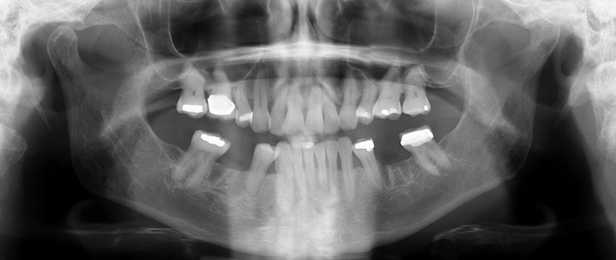
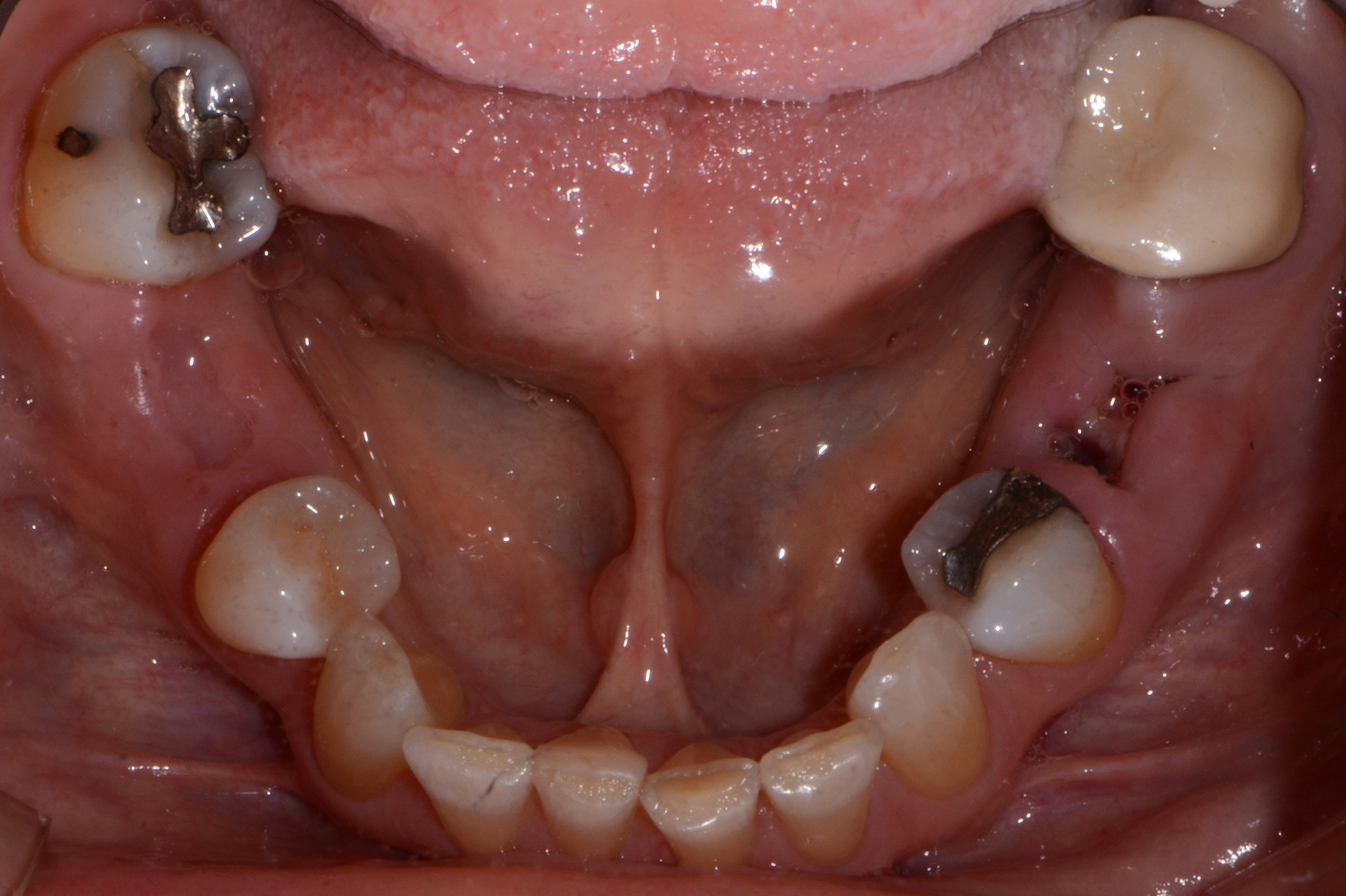
Tooth no. 19 was isolated with a dental dam under local anesthesia. All existing restorative material and decay was excavated leaving a very evident crack into the pulp, down to the pulpal floor. The mesial canals were completely calcified, the decay extended beyond the bone level. This tooth was diagnosed as hopeless and scheduled for removal. A provisional was placed at this appointment.

Tooth no. 30 was deemed hopeless at initial evaluation and extracted.

Phase II: Diagnostic Data Accumulation and Treatment Planning

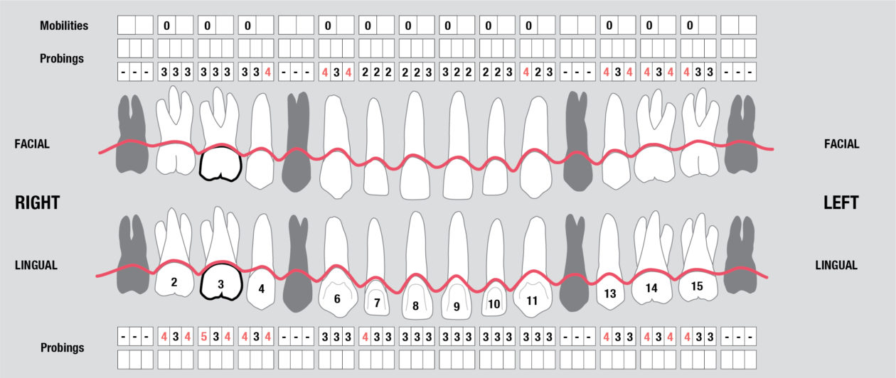
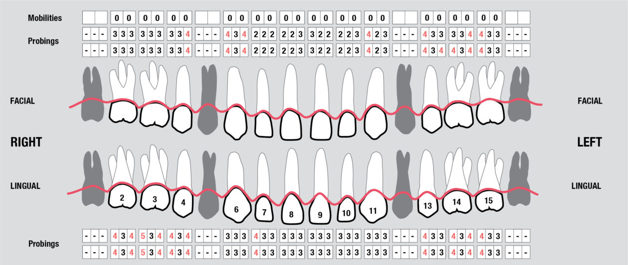
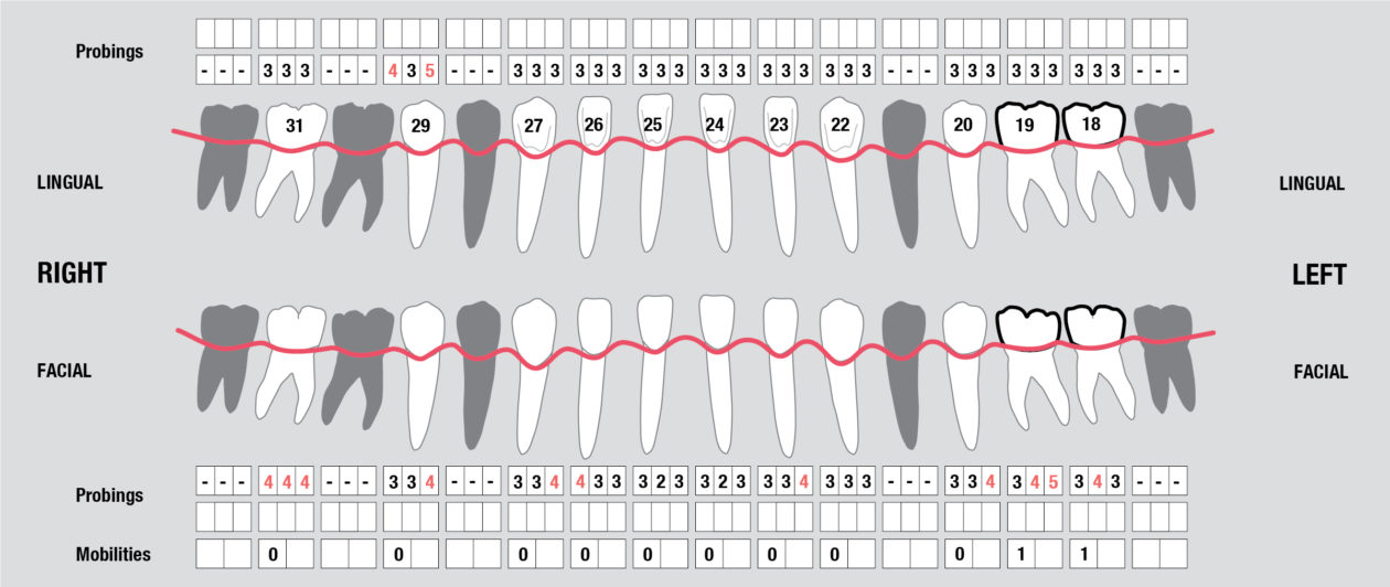
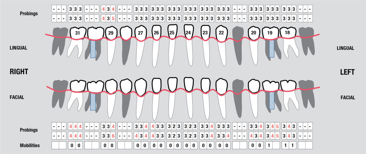
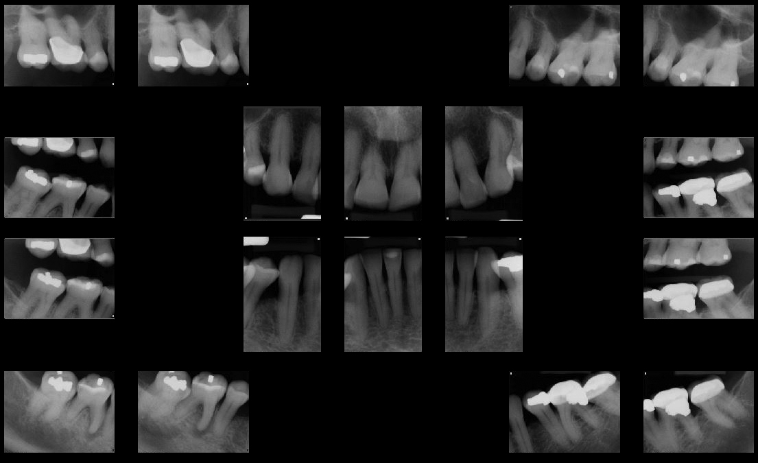
Full Mouth Radiographic Series, Maxillary and Mandibular Intraoral Scans, Digital Jaw Relation Recording with a Leaf Gauge, Complete Photographic Series, Periodontal Charting, Restorative Charting.

Phase III: Treatment Planning Consultation and Treatment Presentation

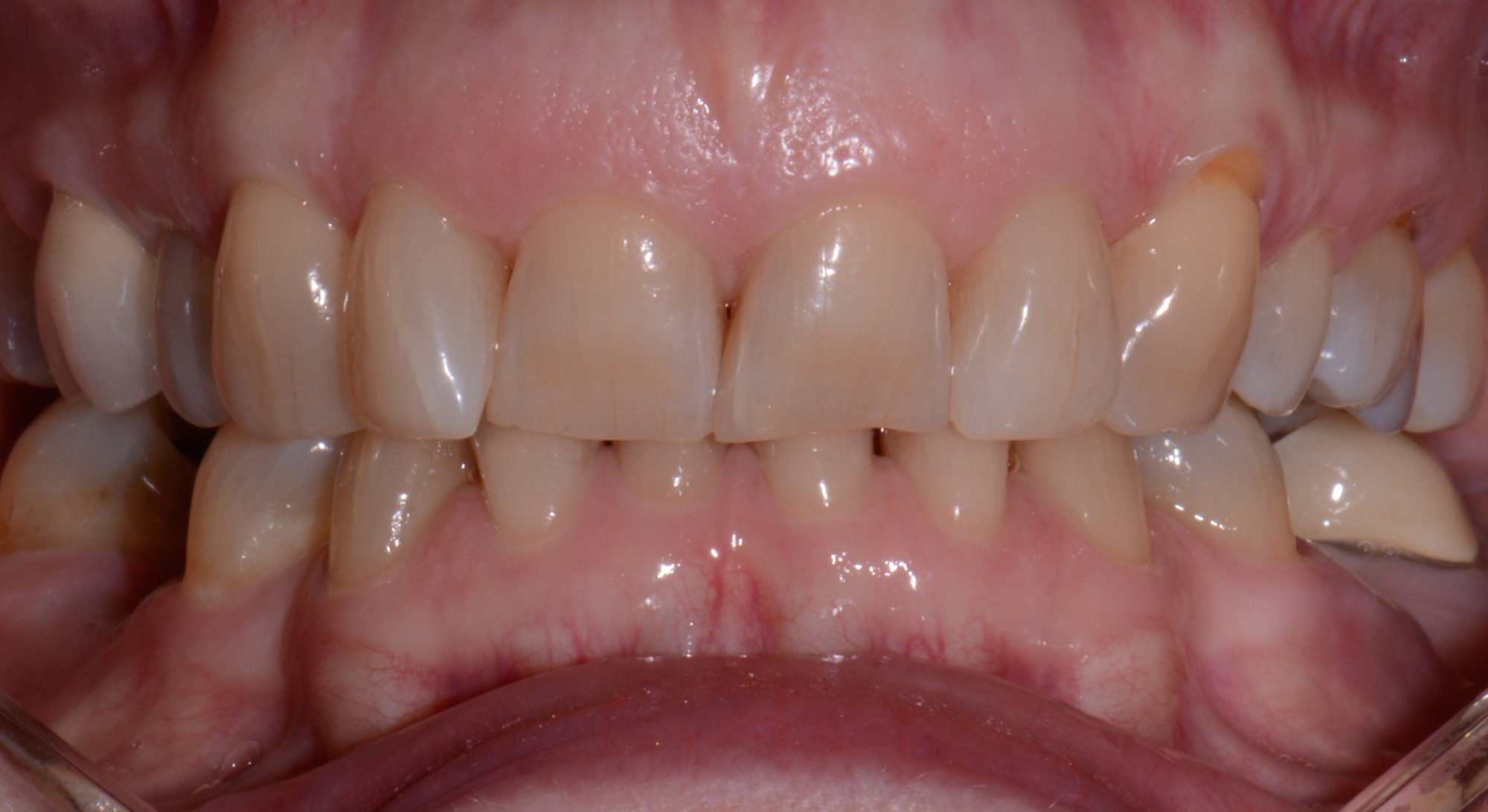
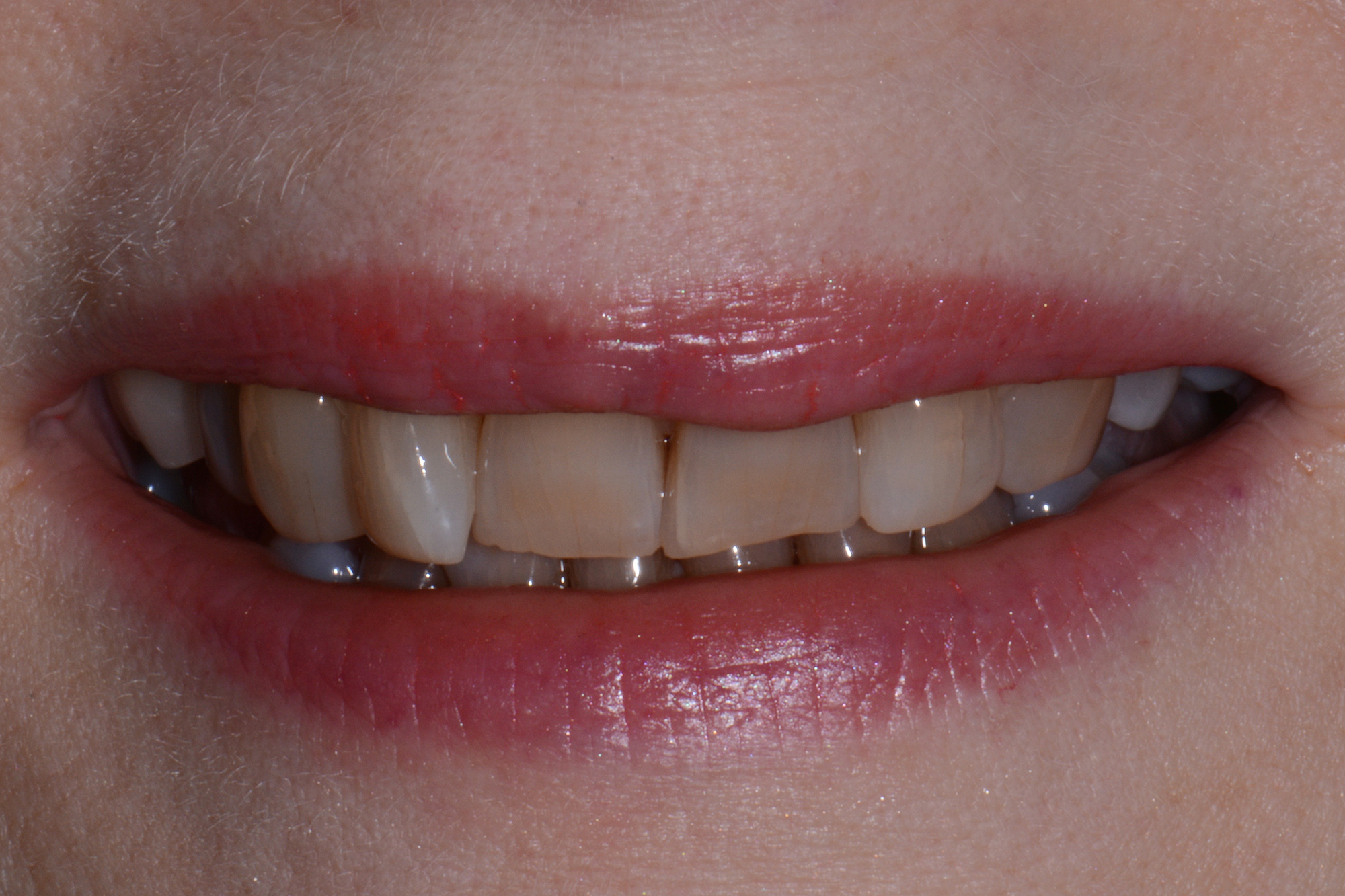
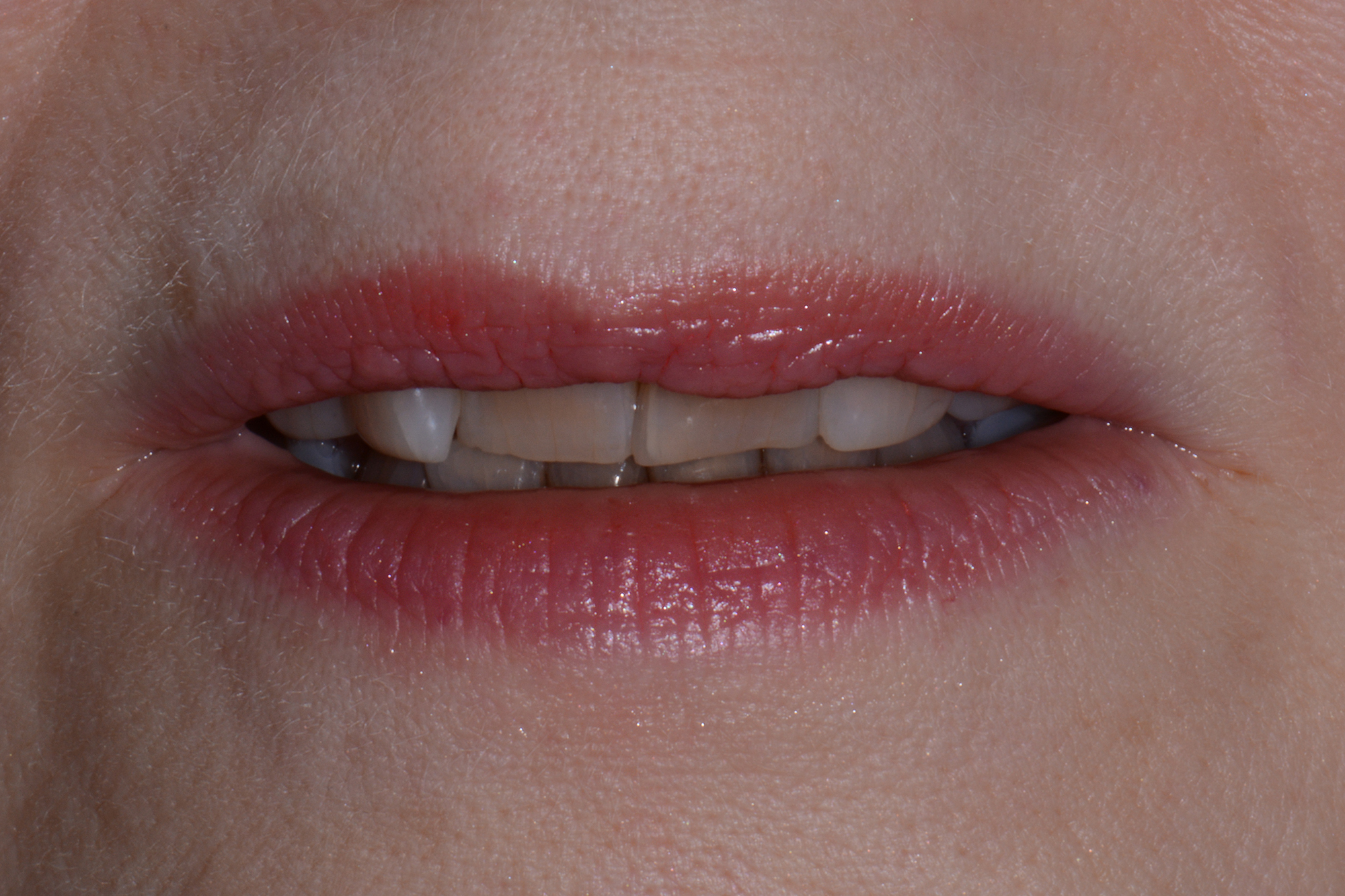
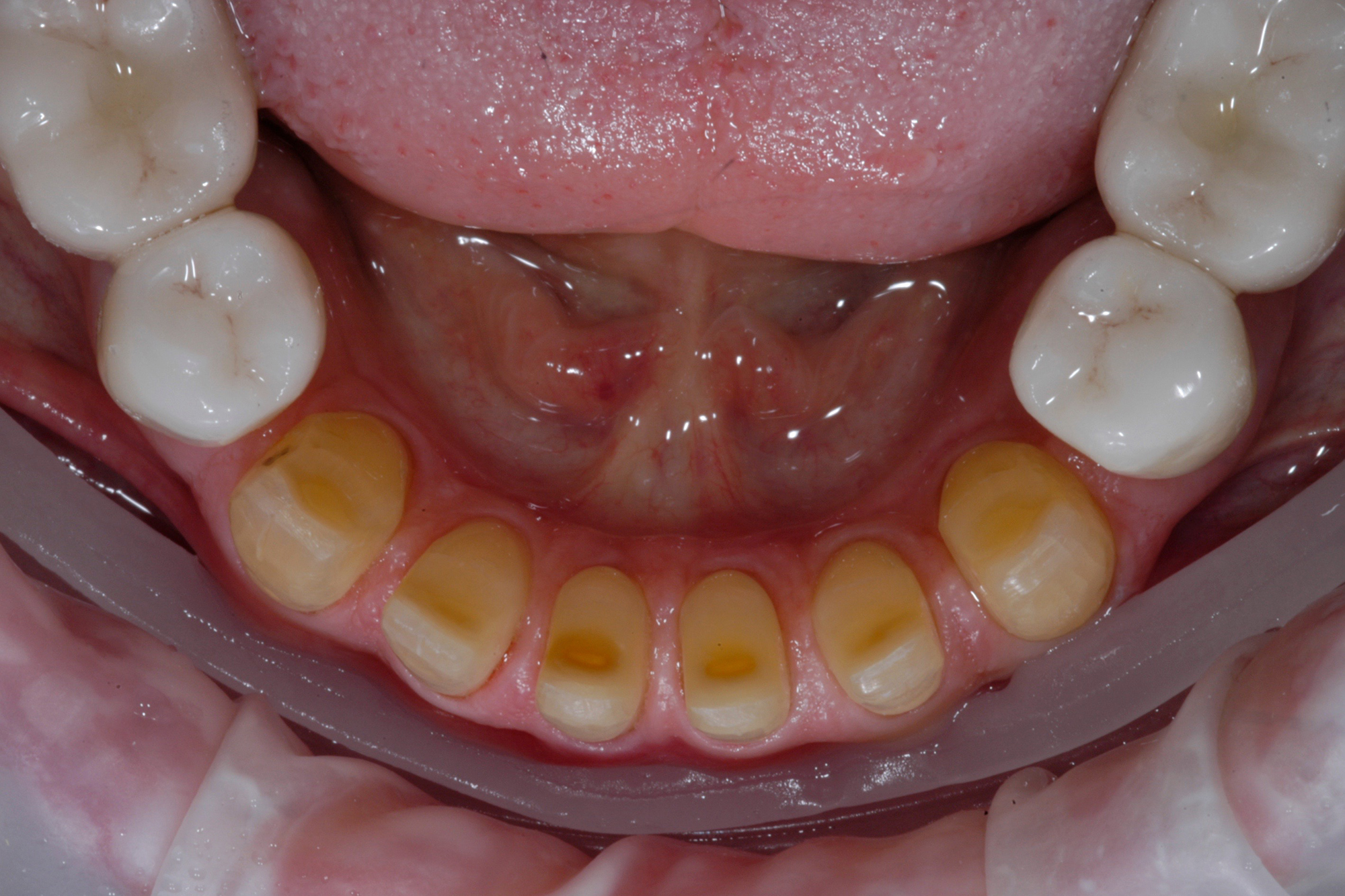
In considering a treatment plan for our patient, it is important to note that in our initial information gathering she was clear she had no esthetic concerns. Further, her desires were single tooth in nature, specifically to replace her missing lower molar teeth. The most challenging aspect of this plan was communicating to the patient what her true problem was and having her accept that the problem, if left untreated, was a challenge to her dental health for life. She must also understand the solution to the problem and to appreciate that comprehensive care with orthodontics and restorative dentistry is to solve a long standing functional and structural problem. Esthetic improvements are a consequence of restorative and structural improvements, not a primary driver in the plan. In our multiple consultation appointments, we allowed the patient to ask as many questions as she needed answered. In short, it took her about six months to overcome the single tooth mindset that she had been taught by her previous dental experiences. Photographs were a significant component in our explanation. They demonstrated the excessive wear resulting in the extractions and were an indicator of what she should expect if she chose only to have the missing lower molars replaced. The orthodontic consultation was a key in her accepting care. An opinion from one of our specialist colleagues, another set of eyes if you will, added support for our recommendations. The plan ultimately accepted by the patient was altered from our originally proposed plan per the patient’s request to only restore the mandibular posterior teeth leaving the mandibular anterior teeth unrestored. With this compromise we still felt we could achieve acceptable anterior function. That altered our overall plan in two ways. First, the orthodontic plan would now not include intrusion of the lower anterior teeth as the incisal edges will dictate tooth position, not the gingival levels. The orthodontist would place the incisal edges of these teeth in ideal position. And second, when restoring the mandibular posteriors after placement of the maxillary restorations, we will not alter the VDO.

Phase IV: Periodontal Surgery Therapy

Connective tissue grafting was accomplished pre orthodontically to cover the root surfaces in teeth nos. 11 and 12.

Phase V: Orthodontic and Surgical Therapy

Orthodontic therapy was initiated in January 2016. At the midpoint of her orthodontic treatment, a CBCT was obtained as well as an intraoral scan (3M Tru Def). These data files were uploaded to the Implant Concierge website with a treatment plan to place implants in the position of teeth nos. 19 and 30. When the Level 2 technician at IC had the initial plan designed, we held a GoToMeeting with the orthodontist, restorative dentist, and the technician present, all in their respective offices.

Implant surgical stent.

At this meeting, the orthodontist was instrumental in planning the implants in a more mesial position in the edentulous spaces. A surgical guide was then fabricated and implants (Straumann) were placed in an ectopic position. Screw retained provisional crowns (Bisacryl) were placed on the implants providing anchorage for the orthodontist to move the anterior sextant mesially to correct the A/P relationship between the arches. Composite resin was gradually removed from the distal of the provisionals and added to the mesial to maintain ideal contours.

Ectopic implant position tooth no. 30 pre orthodontic treatment.
Ectopic implant position tooth no. 19 pre orthodontic treatment.
Tooth no. 30 implant position post-orthodontic treatment.
Tooth no. 19 implant position post-orthodontic treatment.

Phase VI: Maxillary Restorative Therapy

At the conclusion of orthodontic therapy, we obtained new intraoral scans and a jaw relation recording using a leaf gauge. We sent photographs and the IOS to the laboratory (Summit Dental Lab) for digital design of the maxillary restorations. The lower posterior design was completed opposing the newly designed maxillary arch. Digital design enables very efficient review of the plan between the lab technicians and the restorative doctor. Modifications can be communicated easily, and the final plans can be 3D printed. These models were then used for preparation guidance and provisional matrices.

Under oral sedation in a six-hour appointment, the maxillary teeth were prepared as follows. The original leaf gauge jaw relation recording was retained to be used to replicate the preoperative A/P and vertical position as closely as possible. After the anterior teeth were prepared, we used these records to obtain an anterior jaw relation recording. We then prepared the UR and placed the new anterior record and the original left side posterior record in place to capture the right posterior inter arch relationship. We followed with preparation of the left posterior teeth and got the final piece of the inter arch position using the new records previously obtained. Retraction cords were placed (Ultradent) and margins refined. Second cords were placed for lateral retraction and a complete arch IOS was recorded (3M Tru Def). The lower arch was scanned, and provisional restorations were fabricated using matrices from the digitally designed and 3D printed model. A detailed laboratory prescription, face bow recording, and the analog jaw relation recording were then forwarded to the laboratory for fabrication of the final restorations. The provisional maxillary restorations were scanned and forwarded to the laboratory along with the preparation scan. This scan of the provisional restorations was merged with the preparation scan in the laboratory, giving direction in final restoration design and enabling replication of the provisionals that have been accepted by ourselves and the patient.

Digital design again provides efficient interchange between the technician and dentist.

Six weeks later the final restorations were tried in. After confirmation of fit and esthetics, they were bonded in place (3M Rely X Ultimate).

Phase VII: Mandibular Restorative Therapy

Our original plan had been altered by the patient to not include restoring the lower anterior teeth. However, when the patient arrived for the mandibular prep appointment, she informed us that due to the dramatic esthetic improvement of her maxillary teeth she now wanted to restore all the mandibular teeth including the anteriors. The fact that we did not pressure her during the initial consultation visits ultimately led to her choosing ideal care.

After preparing the mandibular posterior teeth this change in plan was accommodated by increasing her VDO arbitrarily, again with a leaf gauge. Scans were sent to the lab for construction of the posterior crowns at this new VDO. The technicians then conventionally waxed the mandibular anteriors to that new VDO.

Six weeks later the final mandibular posterior restorations were tried in and bonded in place (3M Rely X Ultimate). The implant crowns were torqued to 35nCm. Teflon tape and composite resin were used to close the access openings.

Minimal preparation veneers were designed to restore the mandibular anteriors and six weeks later they were bonded in place (3M Rely X Ultimate).

An interocclusal splint was fabricated to serve as a means of protection against parafunction and as an orthodontic retainer.

View in maximum intercuspation post-orthodontic treatment.
Maxillary preparations.
Mandibular posterior preparations.
Mandibular anterior preparations.
Definitive smile.
Definitive smile in maximum intercuspation.
Definitive maxillary arch.
Definitive mandibular arch.
Post-op bitewing.
Post-op bitewing.

Commentary

Definitive full face.

The overall success with this case came about because of our team’s patience in allowing time for the patient’s thinking to shift from single tooth emergency type dentistry to a more comprehensive approach. Too many times we as clinicians follow our natural tendencies to just get going with the dentistry instead of taking the time to allow our patients to evolve into choosing optimum care.

In this case, one primary retrospective consideration is the potential impact that airway restriction/obstruction may have played. While she reported no prior diagnoses at her initial assessment, many clinical observations indicated otherwise. Previous orthodontic therapy with bicuspid extractions, anterior retraction, bruxism, TMJ dysfunction and GERD are all co-diagnoses in airway disease. Our treatment included maxillary and mandibular protraction and increased vertical dimension. Both of these changes increase oral volume and potentially improve air flow. Our patient has had no TM related pain or problems in the 2 years since completion of her care. Future patients with similar situations will be screened for airway disease.

A review of treatment goals indicates complete resolution of structural and functional disease with the expectation of long-term stability.

Dr Brian Schroder is a general dentist practicing in San Antonio, Texas. He is an assistant professor in the AEGD Program at the UT San Antonio Dental School, a member of the Speakers Bureau of the Seattle Study Club, and the director of the Alamo Collaborative Dental Forum Seattle Study Club.

Dr Tyler Ferris is an orthodontist practicing in San Antonio, Texas

Dr Amir Hosseini is a periodontist practicing in San Antonio, Texas and is the director of The South Texas Advanced Restorative Seminars Seattle Study Club.