Case 97: Hopeless but Still Useful Primary Lateral Incisors in the Preservation of Vertical and Buccolingual Bone Dimension

Managing cases in which the maxillary permanent lateral incisors are congenitally missing is always challenging. Do we move the canines into the lateral spaces? Do we move the canines distally to open ideal permanent lateral-sized spaces? This case shows the body’s incredible ability to maintain bone volume in both the vertical and horizontal dimensions by preserving the primary laterals in a case with minimal periodontal attachment to the bone.

Introduction

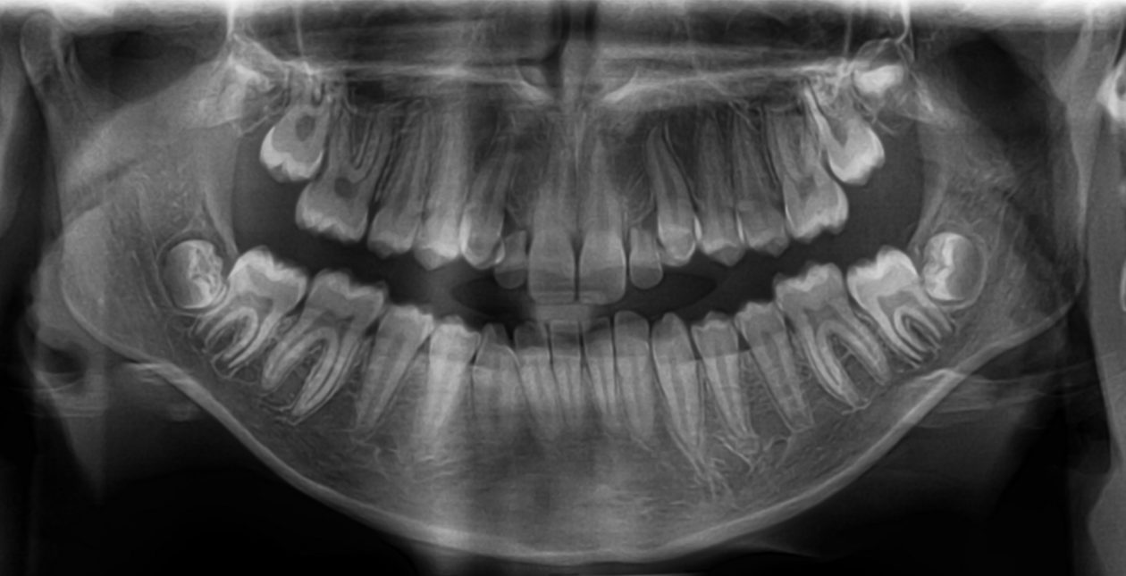
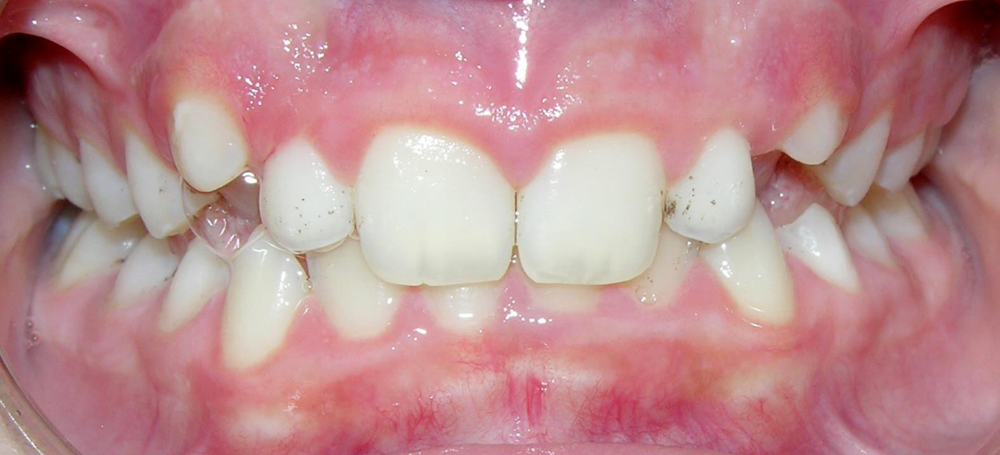
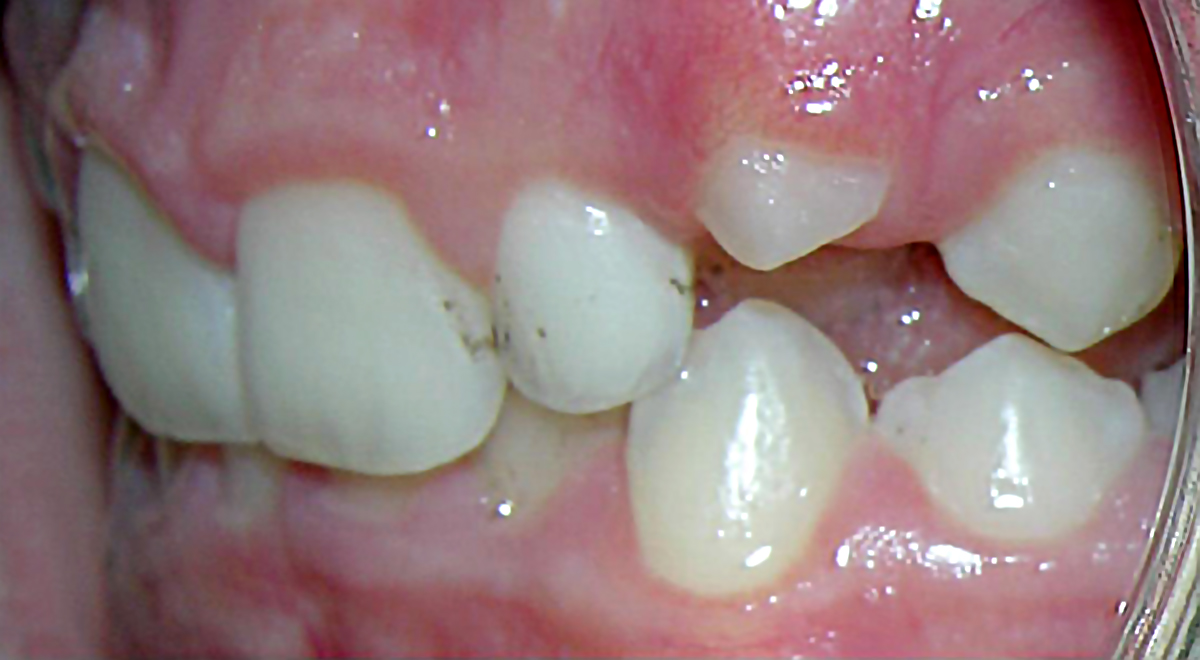
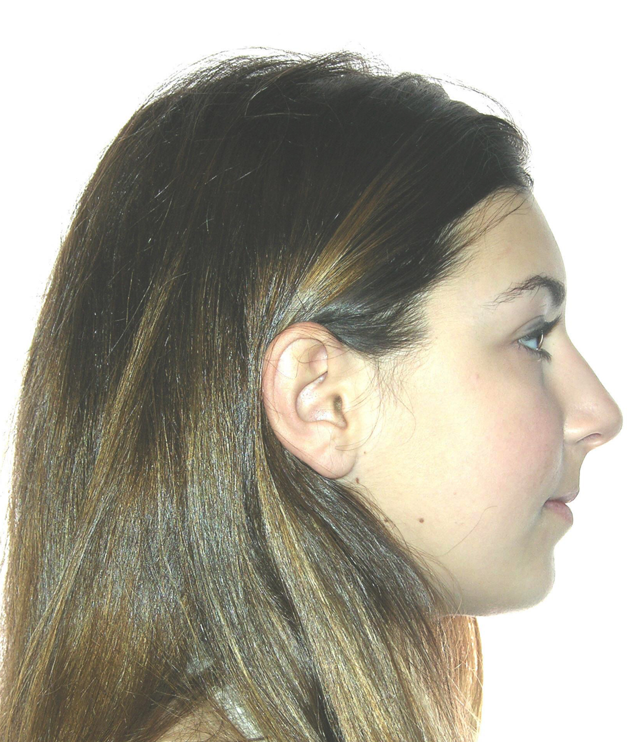
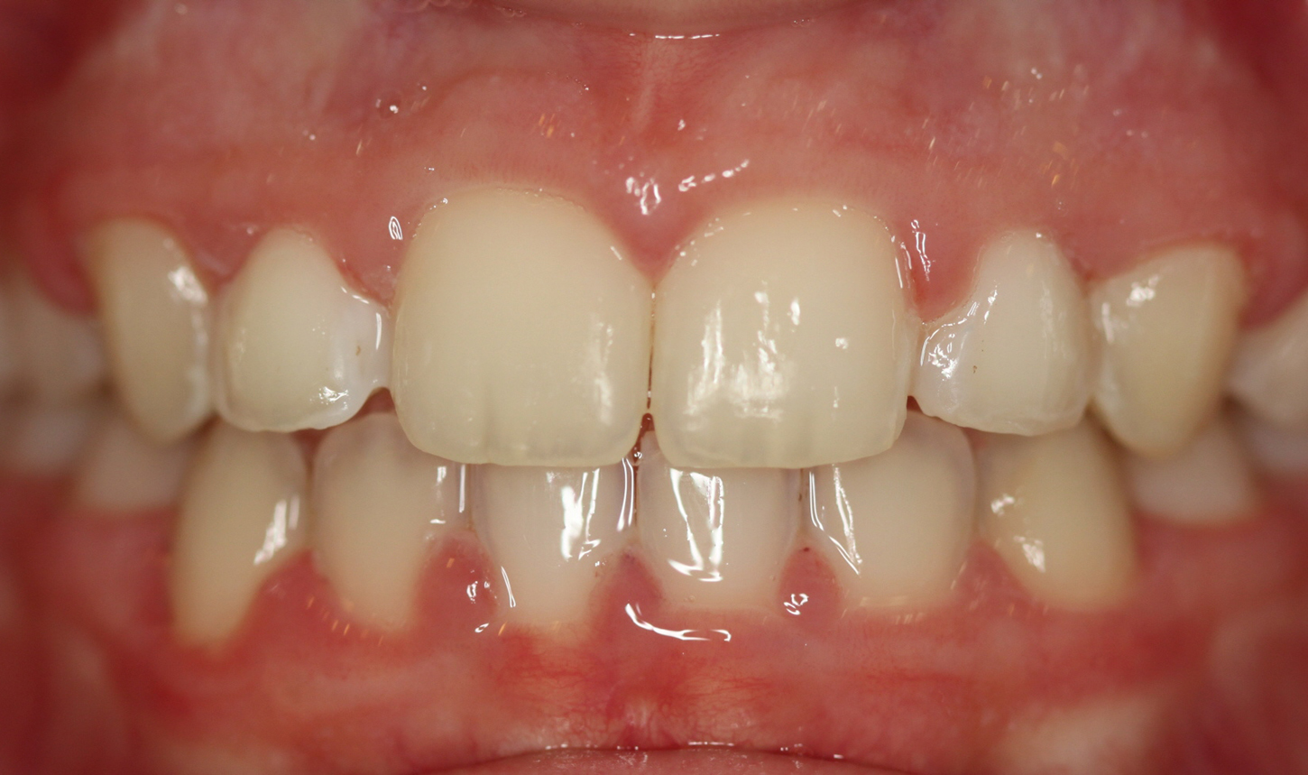
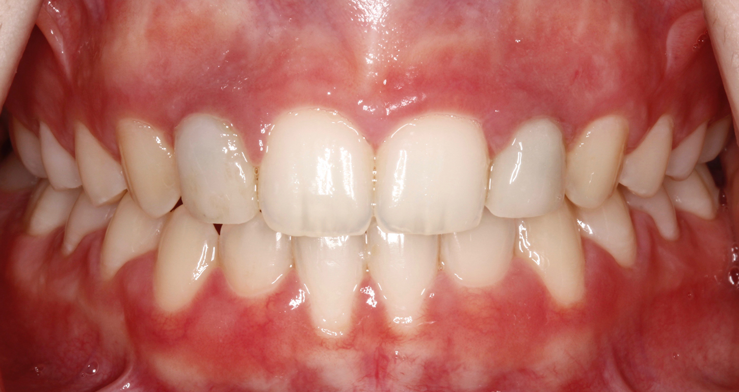
The agenesis of maxillary lateral incisors is a common developmental anomaly which presents an orthodontic and prosthetic challenge that often requires interdisciplinary collaboration for an ideal treatment result. The two most common treatment approaches in these cases are space opening for prosthetic restorations and space closure with the use of permanent canines as substitutes for the missing teeth. In order to meet both esthetic and functional goals, selection of the most appropriate treatment plan requires consideration of the patient’s malocclusion, growth pattern, facial profile and smile line, along with the size, shape and color of the canines.

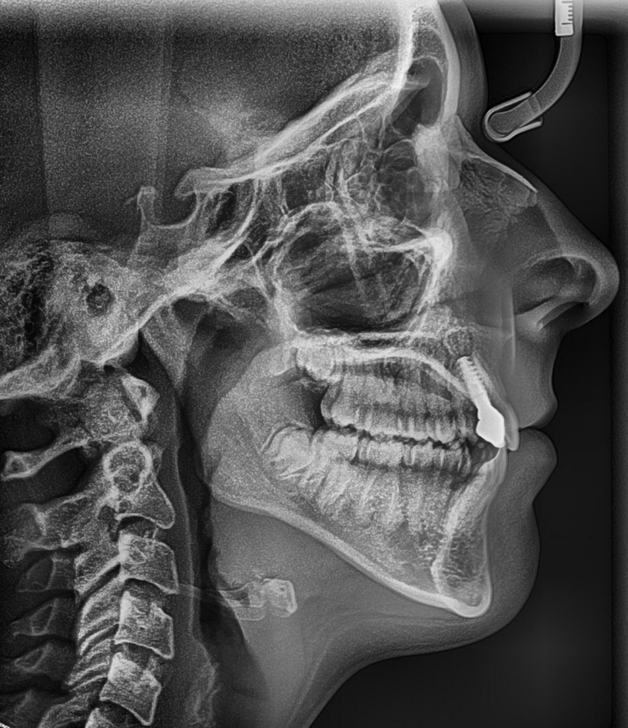
If future placement of single tooth implants is recommended in young patients, special care must be taken because implants cannot be placed until facial growth is complete. The late Dr Vince Kokich suggested that in these cases, the lack of eruption of permanent lateral incisors prevents full development of the osseous ridge needed to place implants. As such, he recommended extraction of the primary lateral incisors in order to encourage eruption of the permanent canines next to the central incisors. He then recommended orthodontic distalization of the canines into their ideal positions in order to allow for the development of the buccolingual width needed for future implant placement.

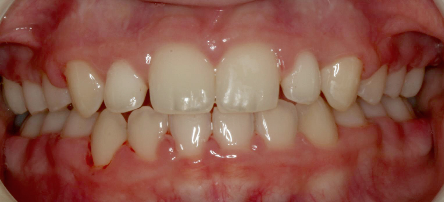
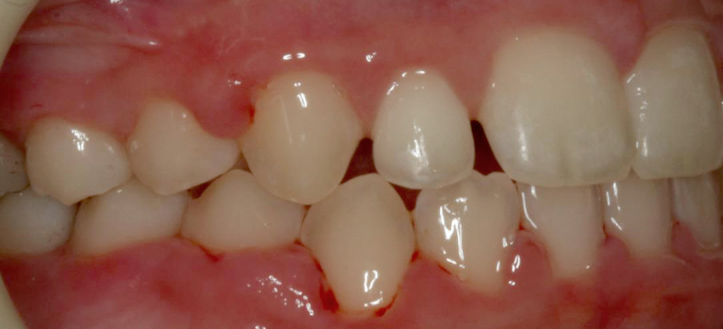
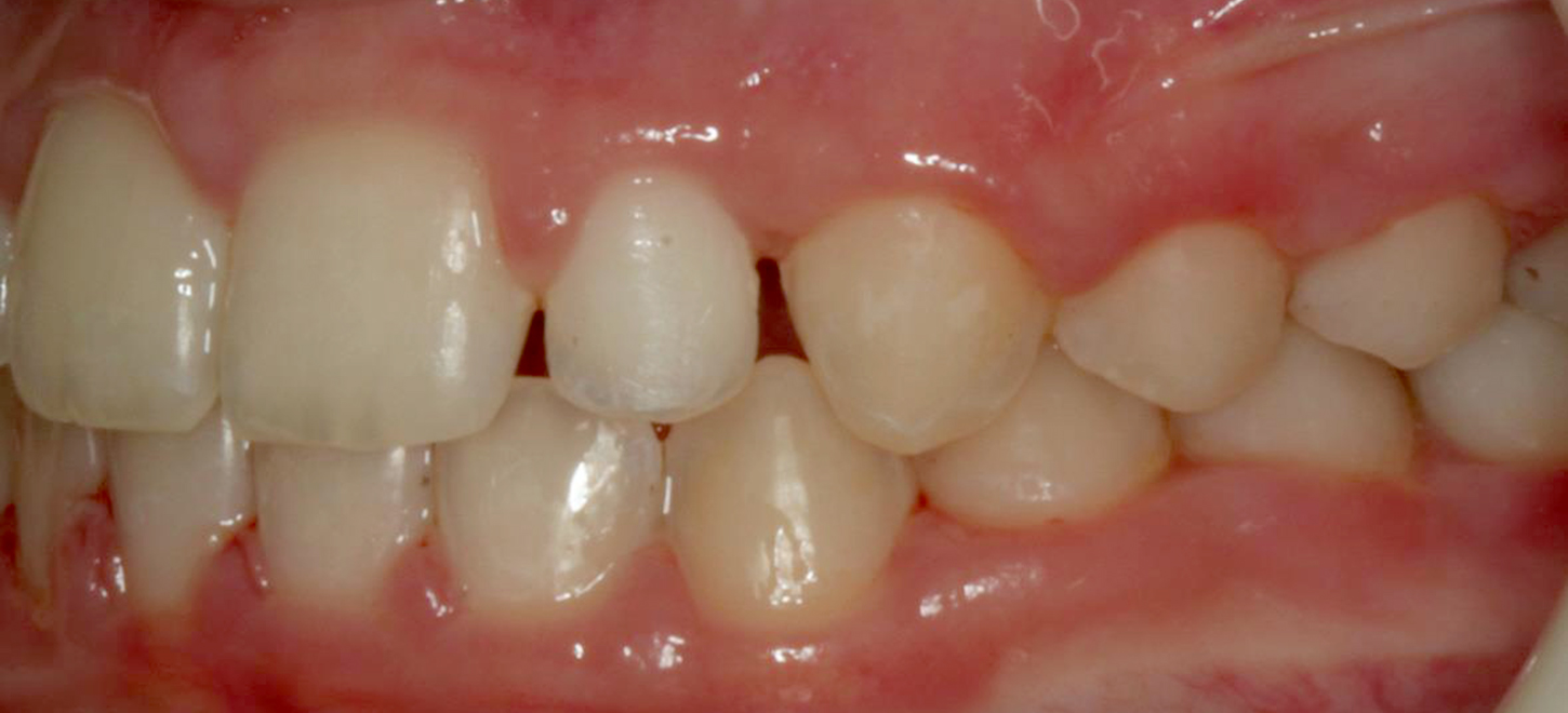
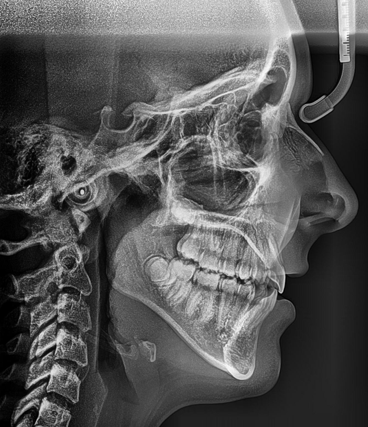
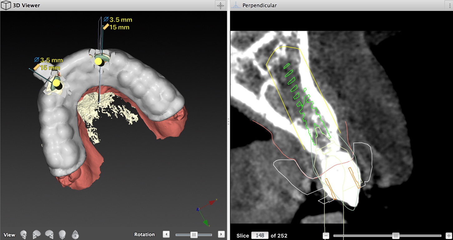
In this case study, we present an alternative approach for the successful maintenance of both vertical and buccolingual bone dimension for future implant placement in a growing patient. Given that bone augmentation tends to be less predictable in the vertical dimension than in the horizontal, it is our opinion that maintenance of vertical bone level is more critical in cases where single tooth implants are planned. Maintenance of vertical bone is particularly important in these cases because its loss can present a cosmetic challenge, especially when patients have high smile lines.

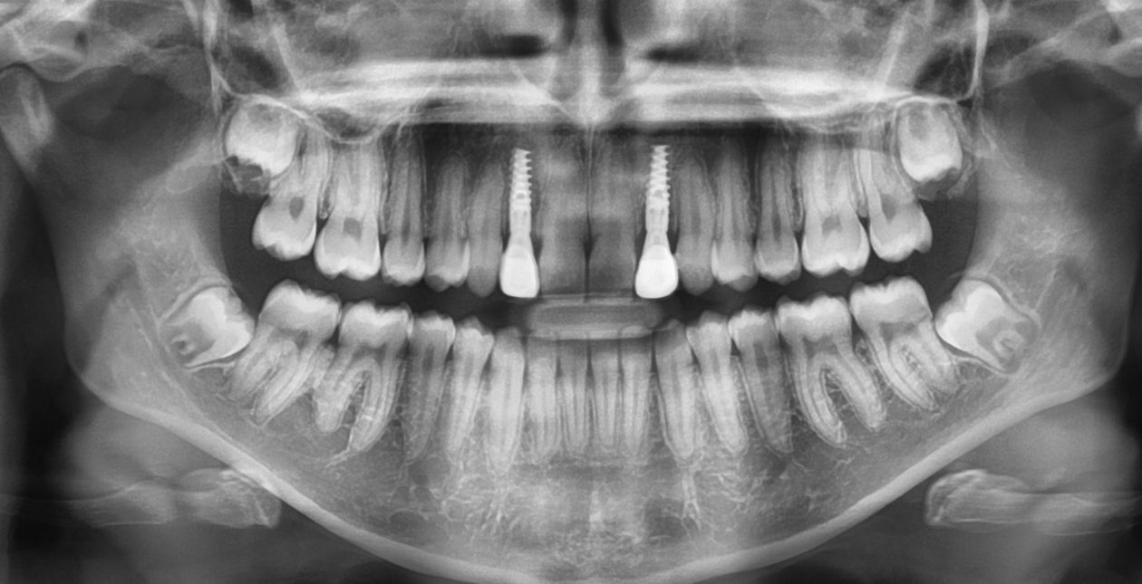
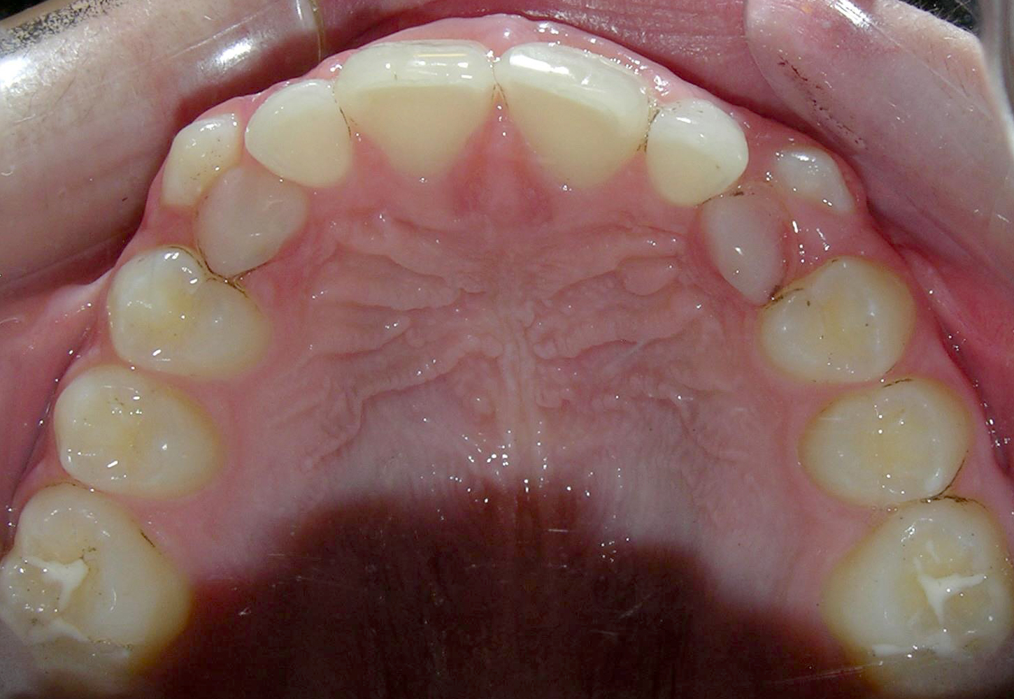
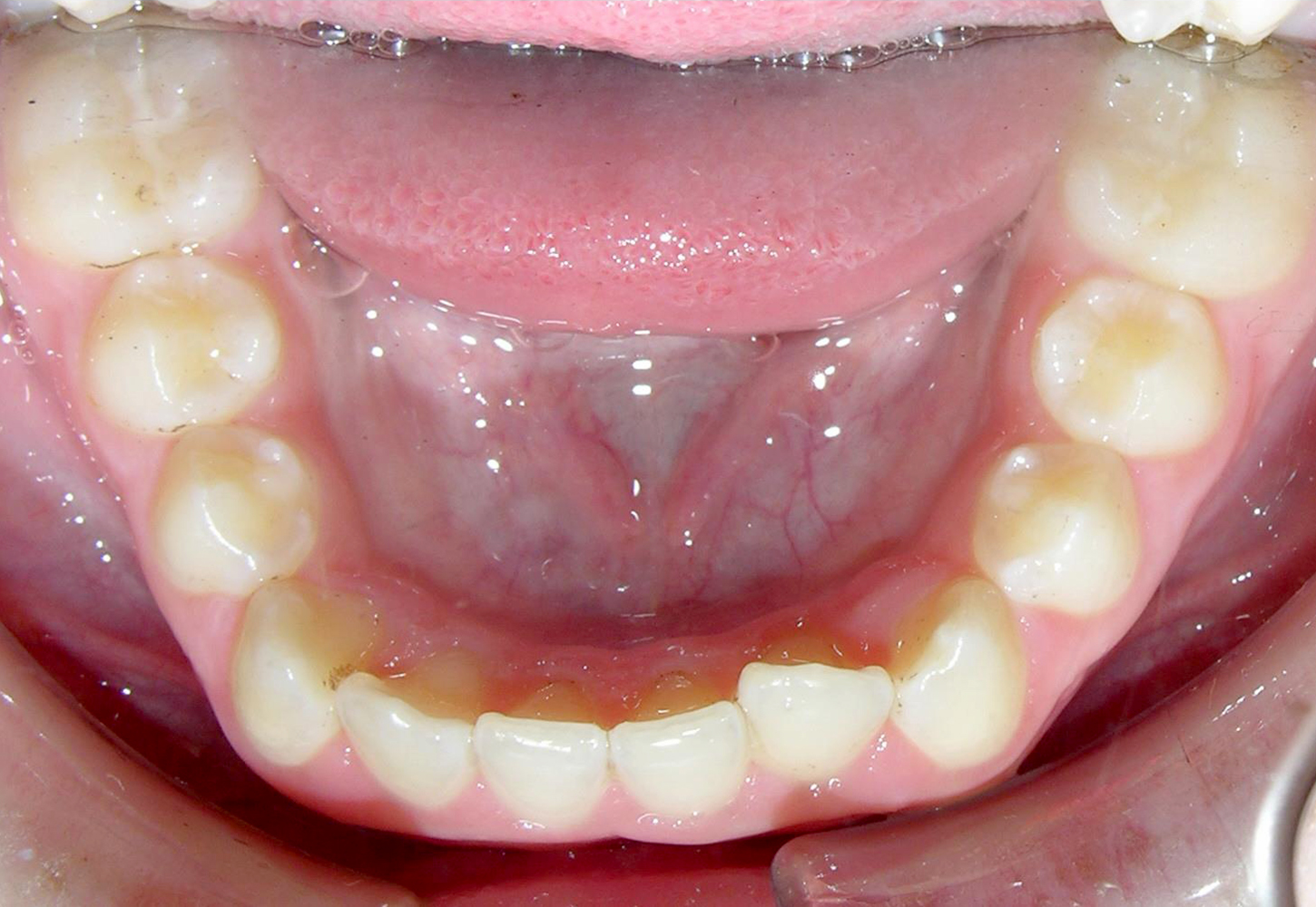
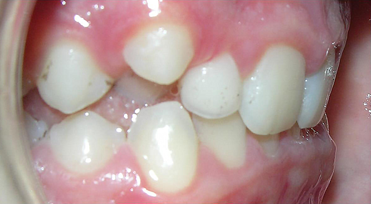
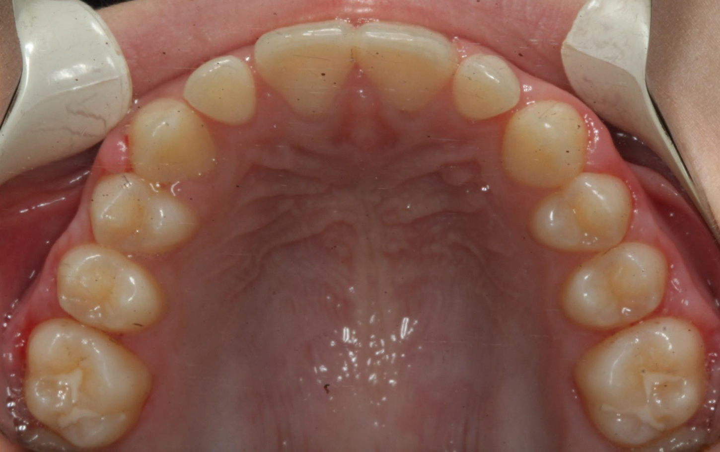
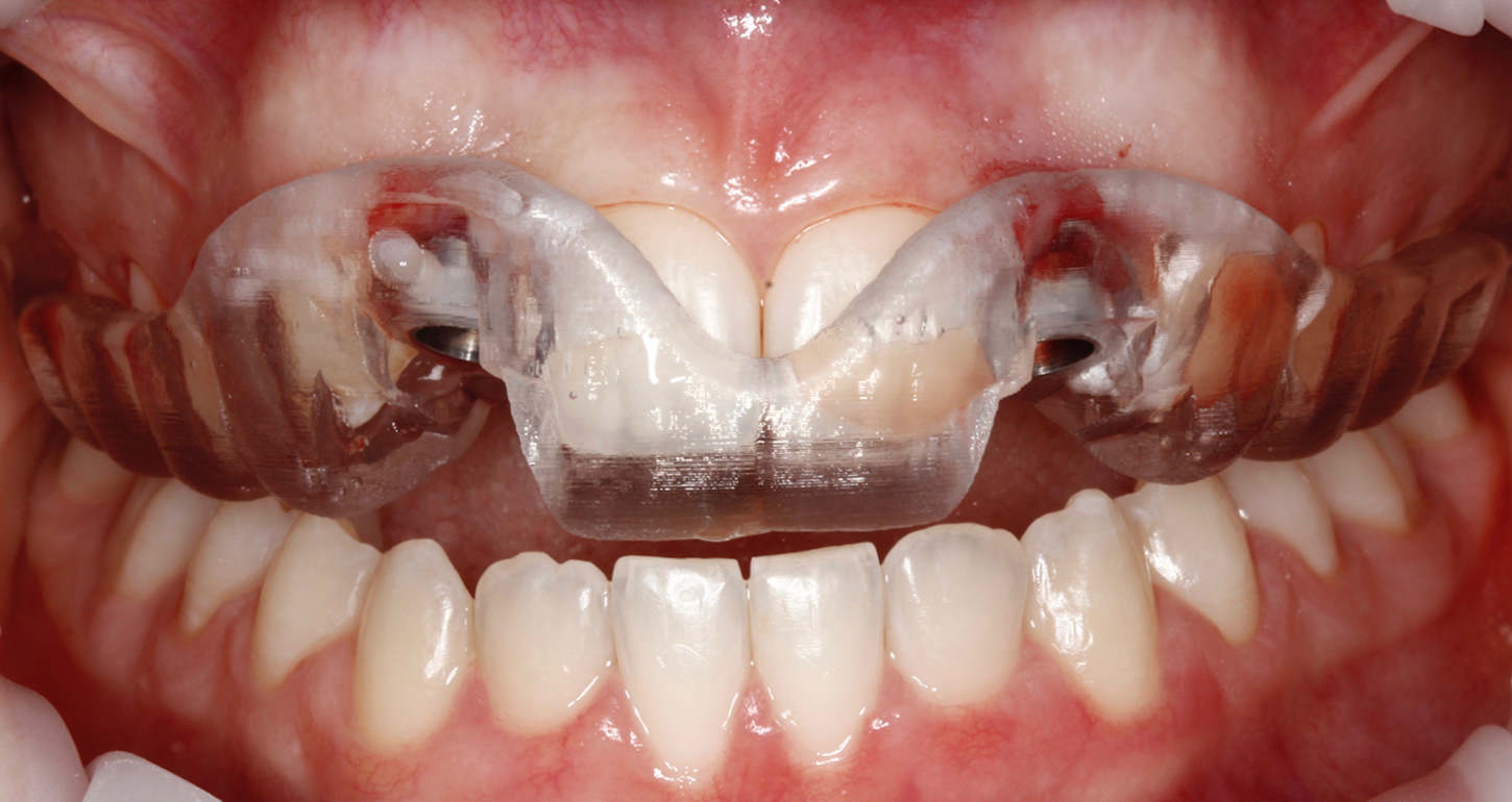
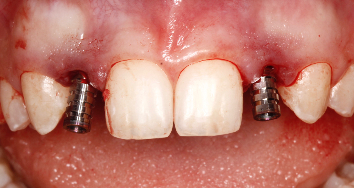
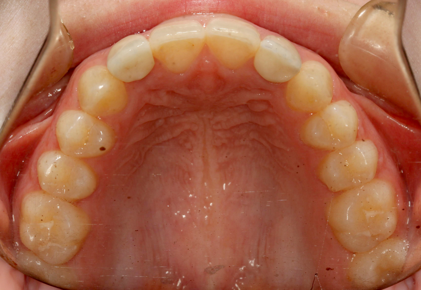
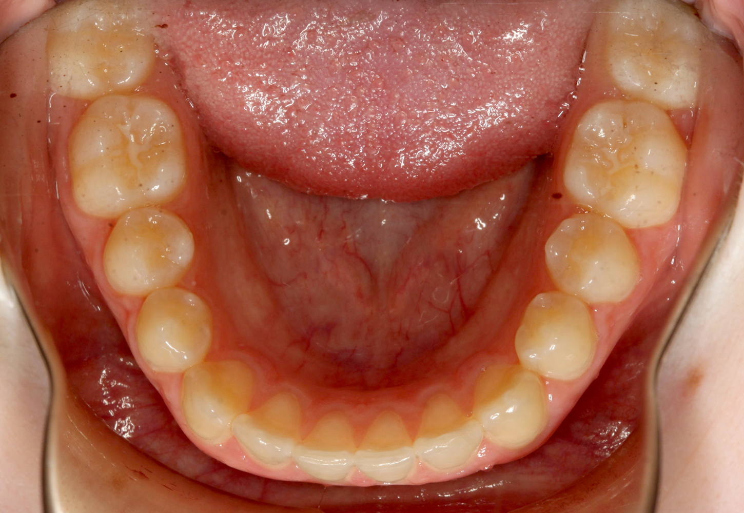
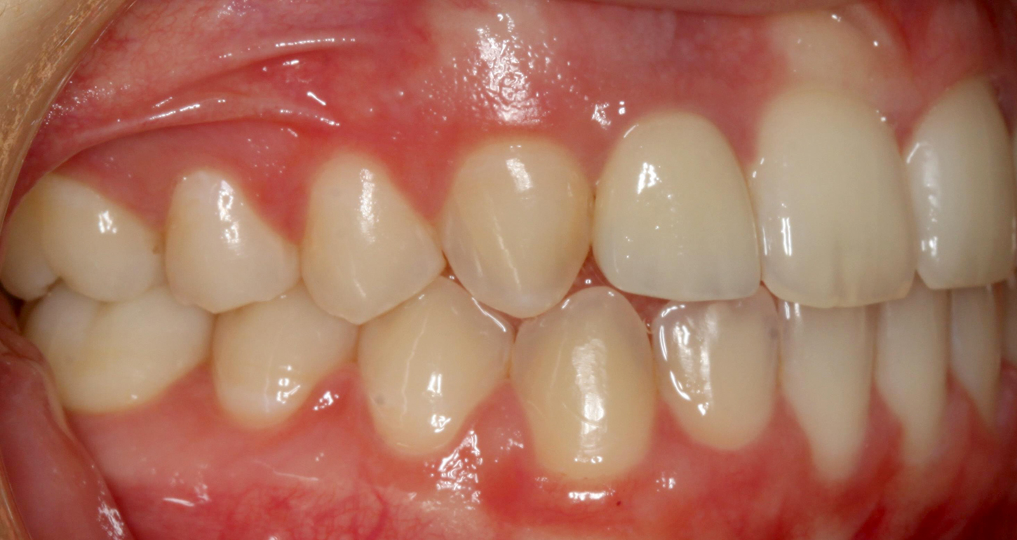
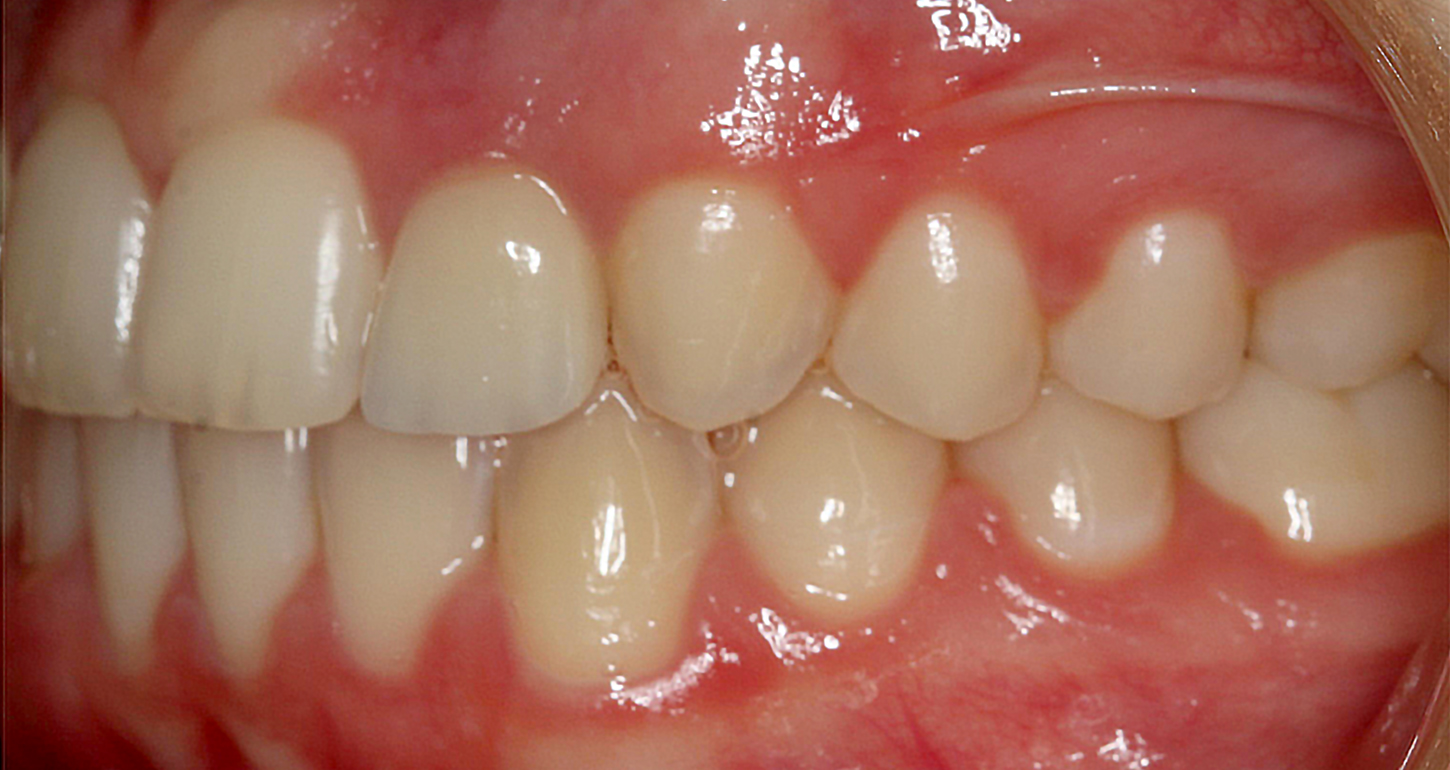
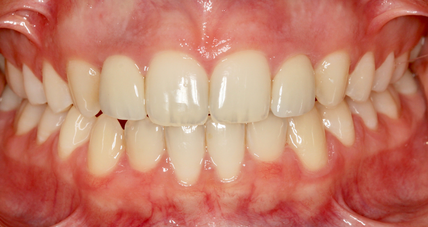
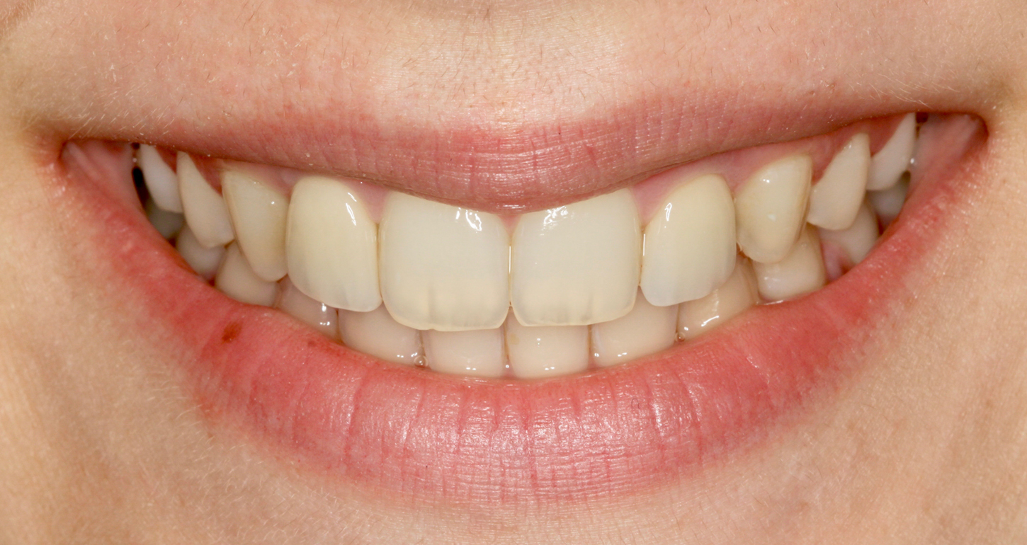
By illustrating a case in which primary maxillary lateral incisors are maintained in a patient whose permanent maxillary laterals are missing, we show successful preservation of both vertical and buccolingual bone dimension during and after orthodontic treatment. We then demonstrate successful placement of single tooth implants without the need for bone augmentation after the completion of growth.

Medical History

The patient was in excellent health. She reported no significant medical history and no allergies. She was not taking any medications.

Initial full face, smiling.
Initial full face in repose.
Initial profile.
Initial lateral cephalometric image.
Initial maxillary occlusal view.
Initial mandibular occlusal view.

Diagnostic Findings

Extraoral/Facial

  • Facial form: Symmetric and balanced.
  • Lower third and middle proportions: Normal.
  • Profile: Convex.
  • Nasolabial angle: Obtuse.

Skeletal

  • Maxillary and mandibular retrognathism.
  • Class I skeletal pattern.

Dental

  • Missing teeth nos. 7 and 10.
  • Over-retained primary teeth nos. C, D, G, and H.
  • Retroclined upper incisors.
  • Upright lower incisors.

Occlusal Notes

  • Anterior deep bite.
  • Deep Curve of Spee.
  • Canine relationship: Class II right and left.
  • Angle molar classification: Class II division II malocclusion right and left.

Periodontal

  • Mobility: Normal for all teeth.
  • Root coverage: Normal for all teeth.
  • Tissue position relative to the cementoenamel junctions: Normal for all teeth.

Diagnosis and Prognosis

Diagnosis

  • Skeletal Class I pattern.
  • Dental Class II division II malocclusion.
  • Missing teeth nos. 7 and 10.
  • Over-retained primary teeth nos. C, D, G, and H.

Prognosis

  • Good for all permanent teeth.
Initial maximum intercuspation.
Initial right lateral view.
Initial left lateral view.

Summary of Concerns

  • Do we move the canines into the lateral spaces?
  • Do we move the canines distally to open normal permanent lateral-sized spaces?

Active Clinical Treatment

Due to a lack of lip support, canine substitution was not recommended. Closure of spaces on the upper arch would have resulted in further retraction of the upper lip and consequently had a detrimental effect on the patient’s facial profile. Instead, future placement of implants in the maxillary lateral incisor positions was advised. In order to maintain the bone until the patient was ready for implant placement, we recommended preservation of her primary lateral incisors.

Phase I: Comprehensive Orthodontic Treatment

Following full orthodontic banding of the upper and lower arches, leveling and aligning was completed. Space was then created mesial and distal to the primary laterals in order to preserve room for bonding of these teeth to their ideal size and for maintenance of the ideal space needed for future prosthetic restorations. After 23 months, the patient’s orthodontic treatment was completed. A Class I molar and canine occlusion was successfully established.

Intraoral view at deband appointment.
Right lateral view at deband appointment.
Left lateral view at deband appointment.
Full face, smiling, at deband appointment.
Full face in repose at deband appointment.
Profile at deband appointment.
Lateral cephalometric image at deband appointment.
Maxillary occlusal view at deband appointment.
Mandibular occlusal view at deband appointment.

Phase II: Splinting of Primary Laterals

On the same day that the orthodontic appliances were removed, the patient’s primary lateral incisors were splinted to her central incisors in order to maintain space and to allow for stability. The primary laterals were kept out of occlusion in order to minimize occlusal trauma to these teeth.

Full face after splinting of primary lateral incisors.
After splinting of primary lateral incisors.

Phase III: Implant Placement and Immediate Temporization

Once the treating clinicians confirmed that the patient’s facial growth was complete at age 18 using serial lateral cephalometric radiographs, implants were placed in the upper lateral incisor positions and subsequently restored. Implant placement was digitally treatment planned in order to allow for guided placement. NobelActive NP 3.5 x 13 mm implants were placed in both implant sites.

Immediate temporization was then completed using Nobel’s TempShell technique.

Digital treatment plan for implant placement illustrating sufficient bone for placement.
Digitally fabricated implant stent to allow for guided implant placement.
Temporary cylinders used for provisional fabrication.
Temporary implant restorations fabricated using Nobel’s TempShell technique.

Phase IV: Definitive Restorative Treatment

Final implant restorations were placed after four months in order to allow for implant integration and soft tissue healing. Zirconia abutments and zirconia crowns (shade A2) were used to achieve ideal aesthetics.

Definitive full face, smiling.
Definitive full face in repose.
Definitive profile.
Definitive cephalometric image of the patient after placement of implants.
Definitive maxillary view.
Definitive mandibular view.
Definitive right lateral view.
Definitive left lateral view.
Definitive implant restorations of the upper lateral incisors.
Up close smile with definitive implant restorations.

Phase V: Maintenance and Retention

After delivery of her final restorations, the patient was given a new upper Essix retainer. The patient was then seen a year following the placement of her implants. She displayed satisfactory maintenance of her final restorations and occlusion.

Commentary

This case report illustrates successful maintenance of both vertical and buccolingual bone dimension in a growing patient whose maxillary lateral incisors were congenitally missing. By retaining the patient’s primary maxillary lateral incisors until the completion of her facial growth, we were able to maintain bone in both dimensions and successfully place implants without the need for bone augmentation. This case highlights the importance of maintaining the primary lateral incisors, regardless of root length and periodontal attachment, in cases where permanent maxillary lateral incisors are congenitally missing.

Dr Patrick T. Cuozzo practices orthodontics in Lincroft and Sea Girt, New Jersey and is the orthodontic advisor to Jersey Coast Dental Forum.

Dr Mehreen Merchant practices orthodontics in Lincroft and Sea Girt, New Jersey.

Dr Anthony M. DiCesare practices periodontics in Red Bank, New Jersey and is the director of Jersey Coast Dental Forum.

Dr Lisa Indelicato practices restorative dentistry in Middletown, New Jersey and is a member of Jersey Coast Dental Forum.