Case 103: Save or extract? – A Case Quandry in Meeting Functional and Esthetic Needs

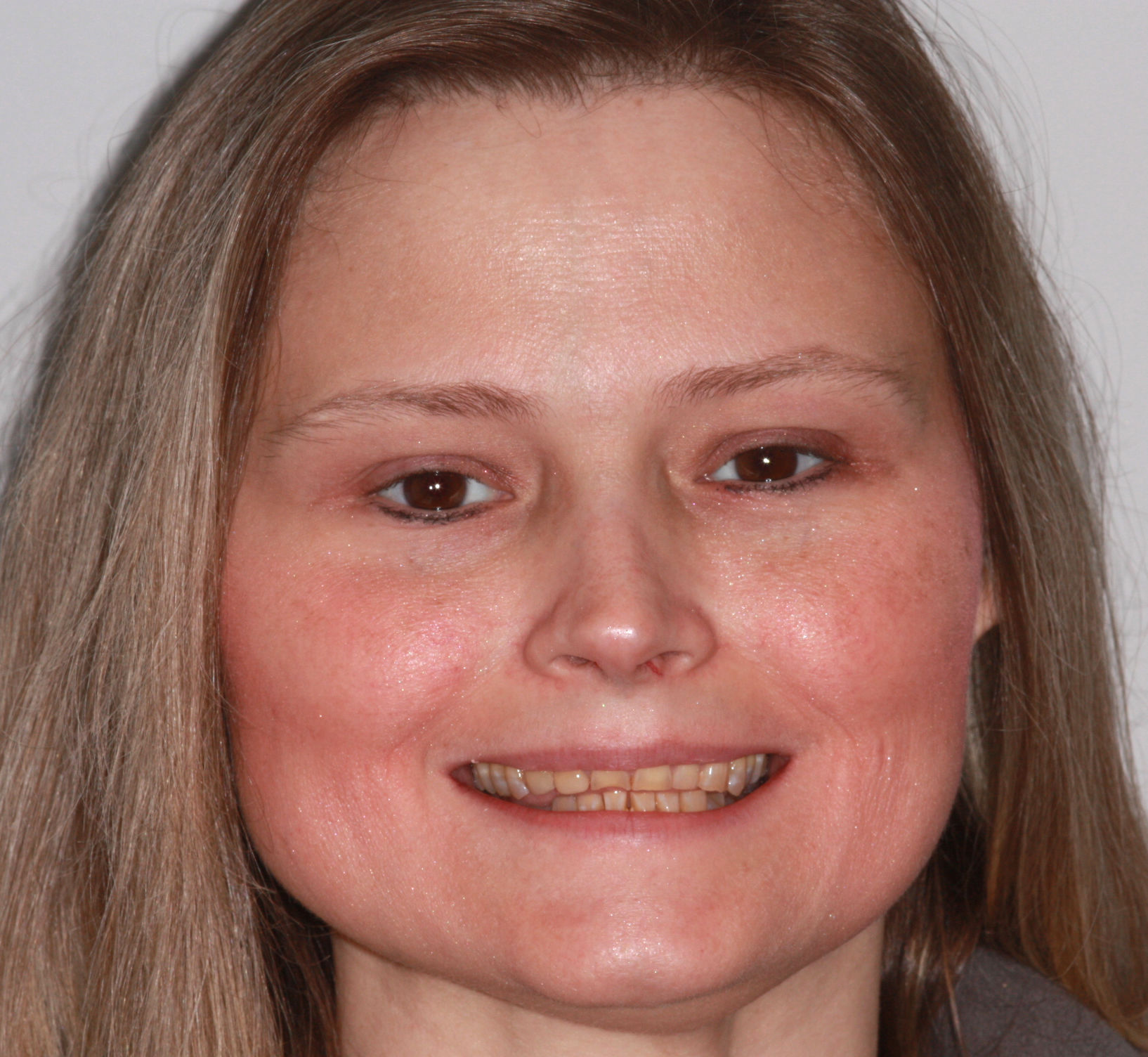
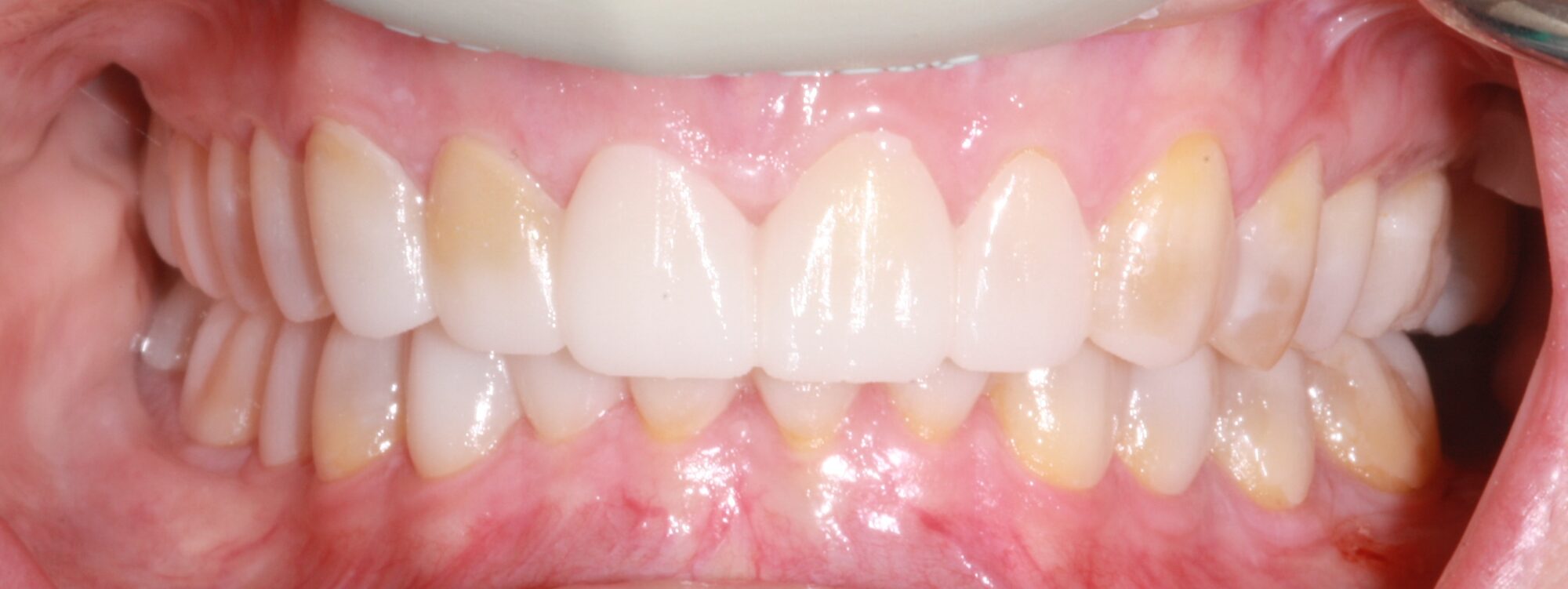
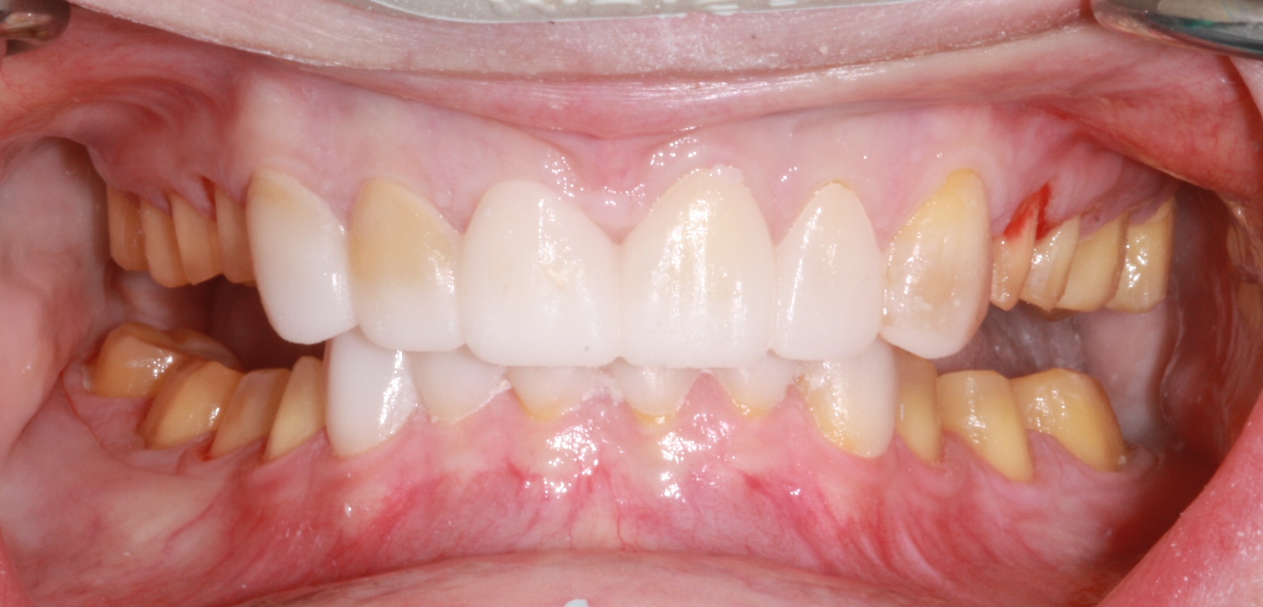
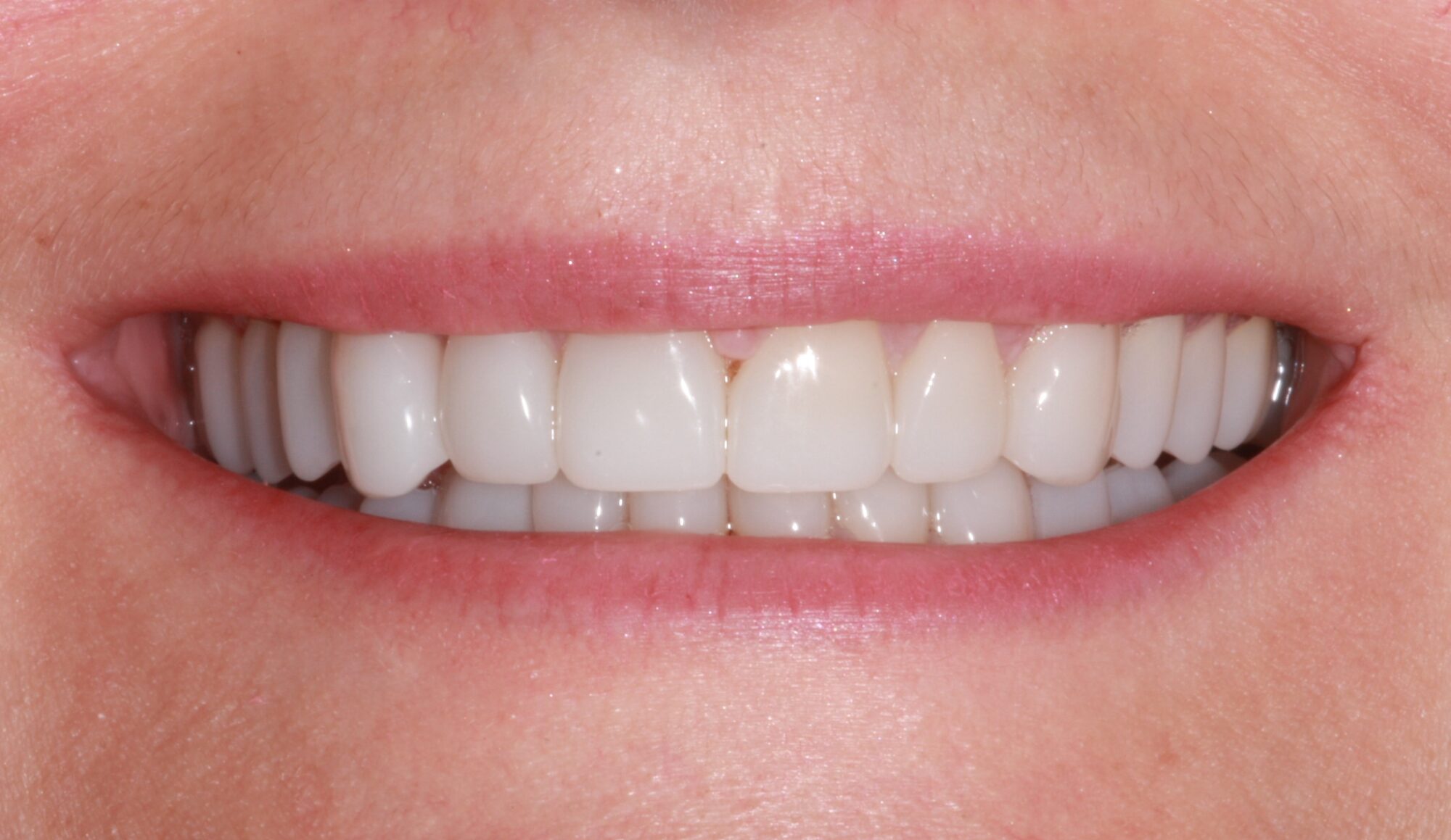
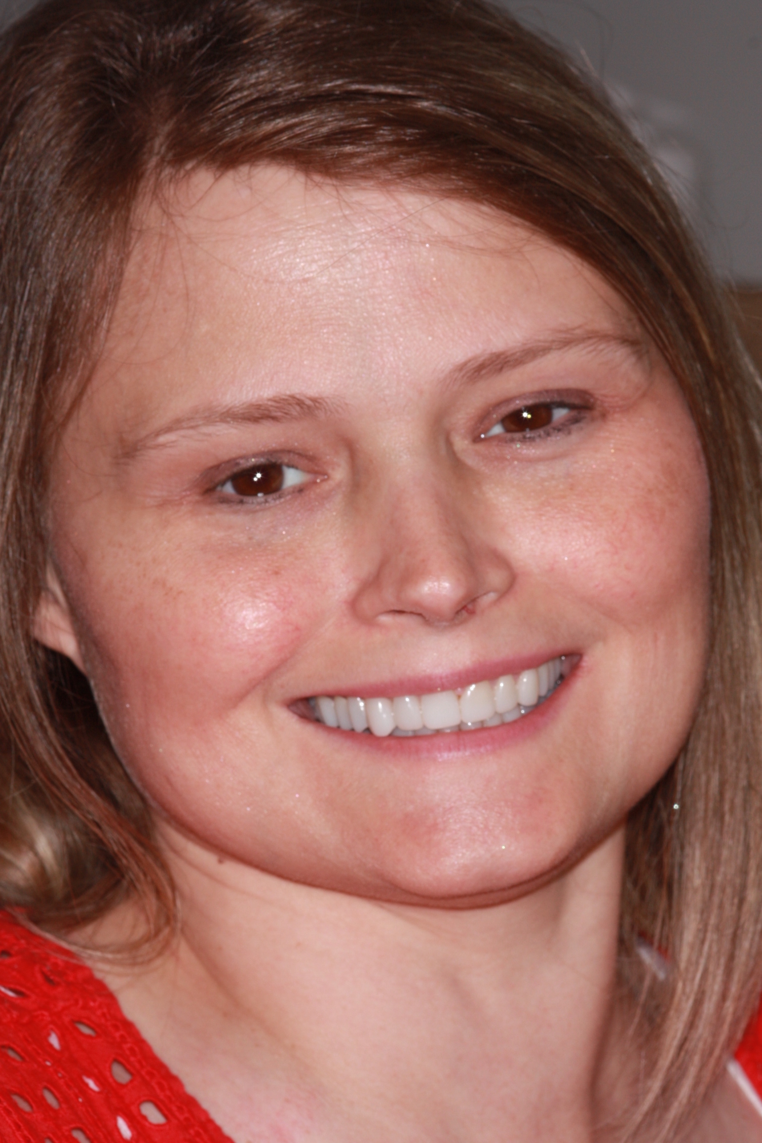
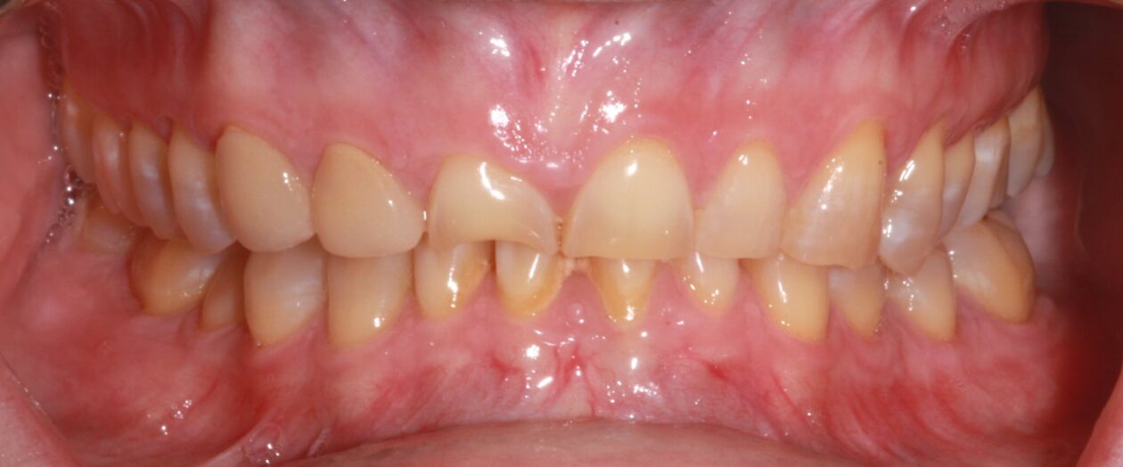
The patient presented to our office seeking a 2nd opinion for treatment options related to treating a deteriorating dentition. She also had concerns about her smile and the appearance of her teeth. She was acutely aware of her asymmetric smile and felt that she looked far older than her age (Fig 1).

Her previous dentist had recommended full-mouth extractions, followed by an implant-supported prosthesis. Although she was willing to accept this, she did not want to lose her natural teeth, if possible.

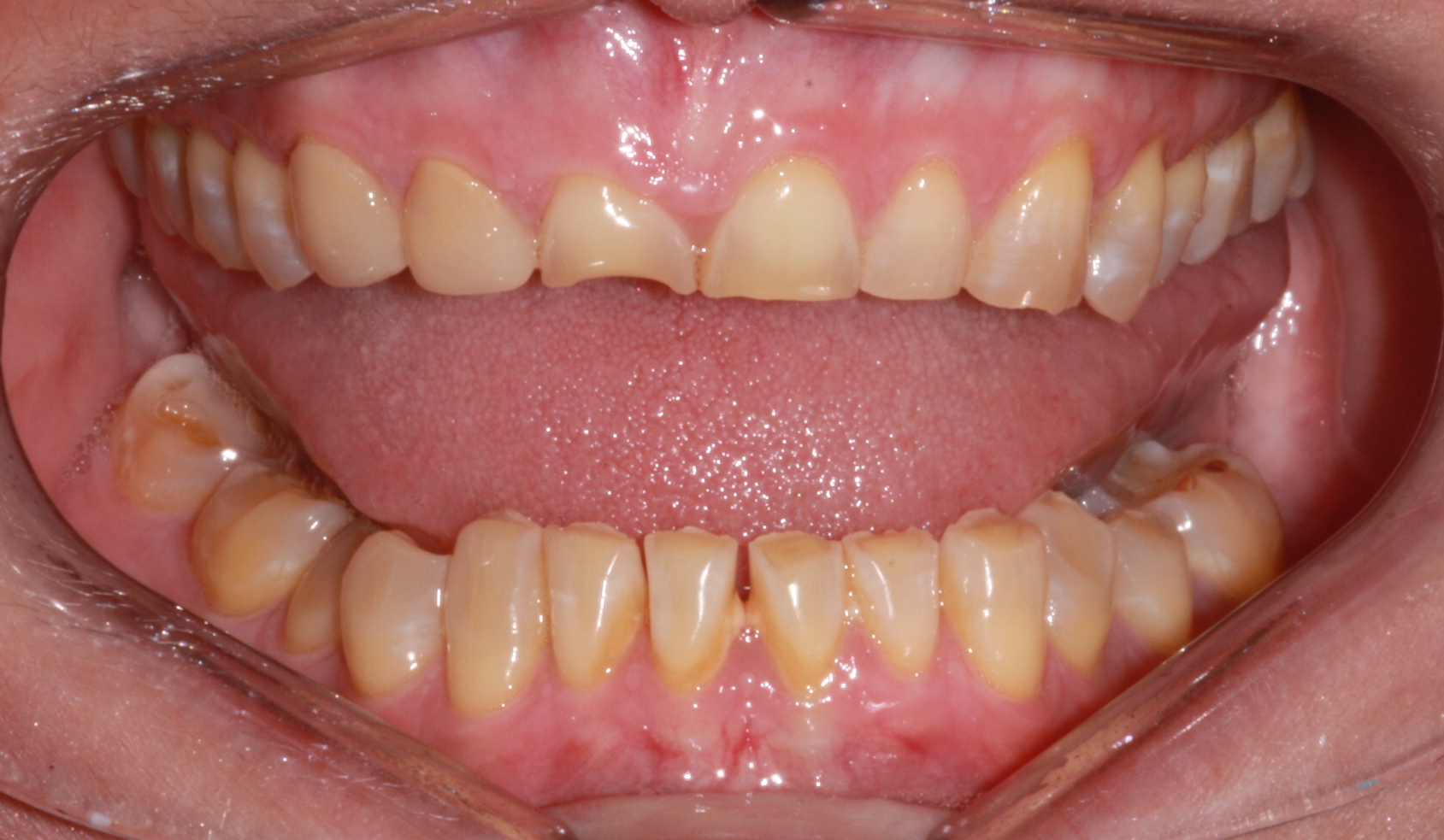
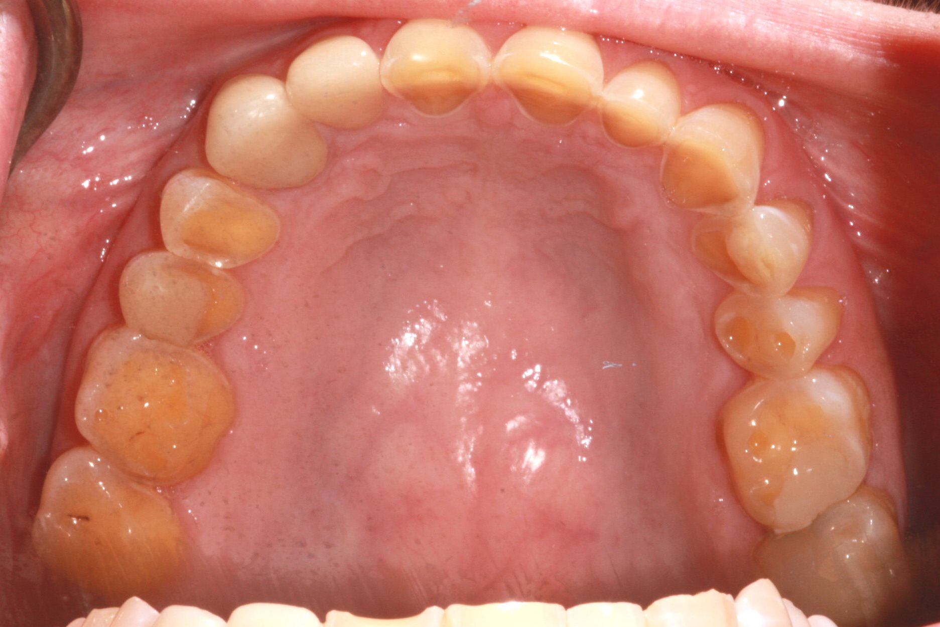
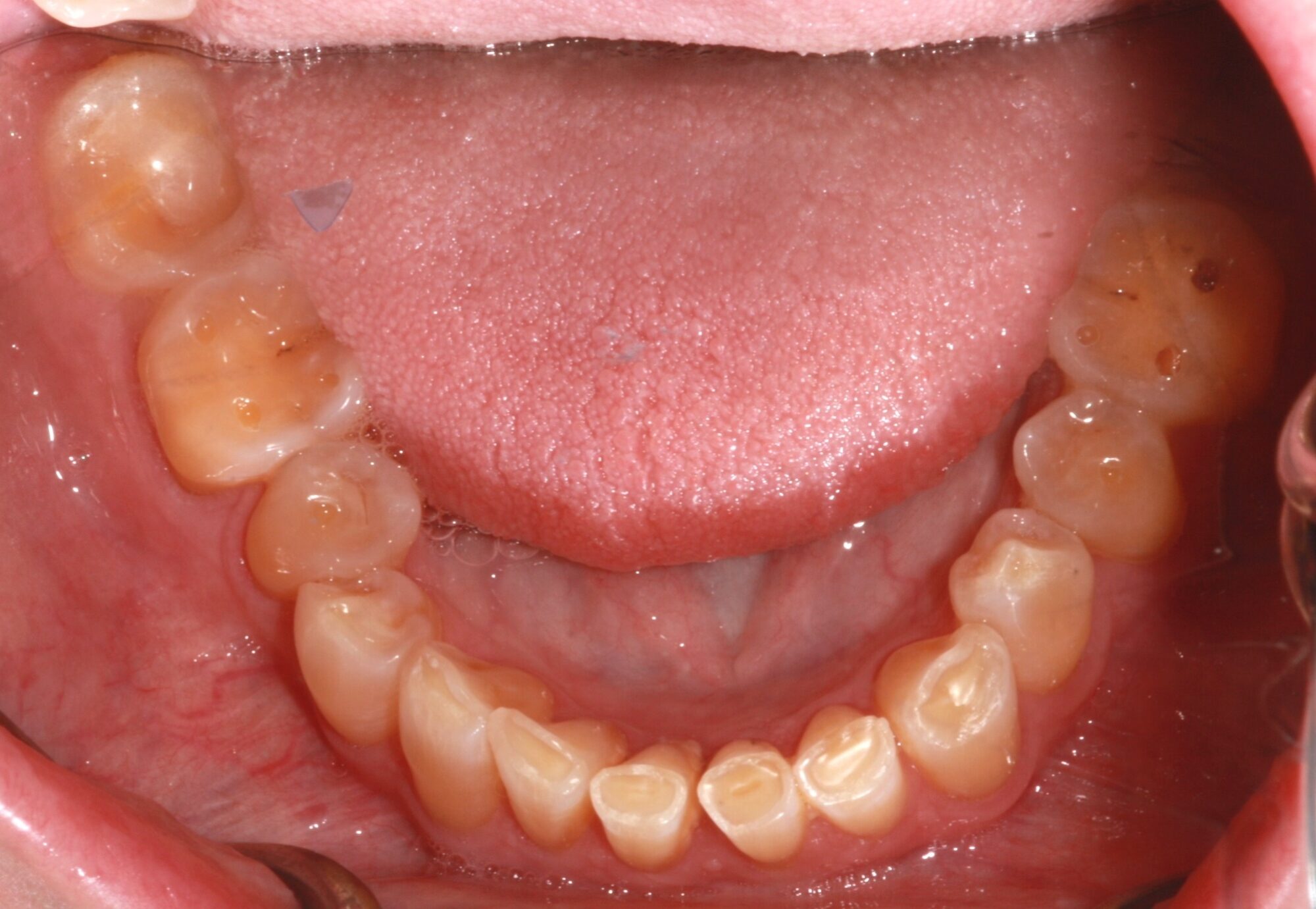
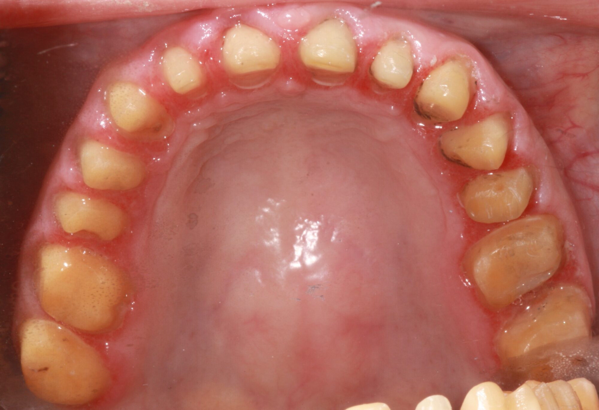
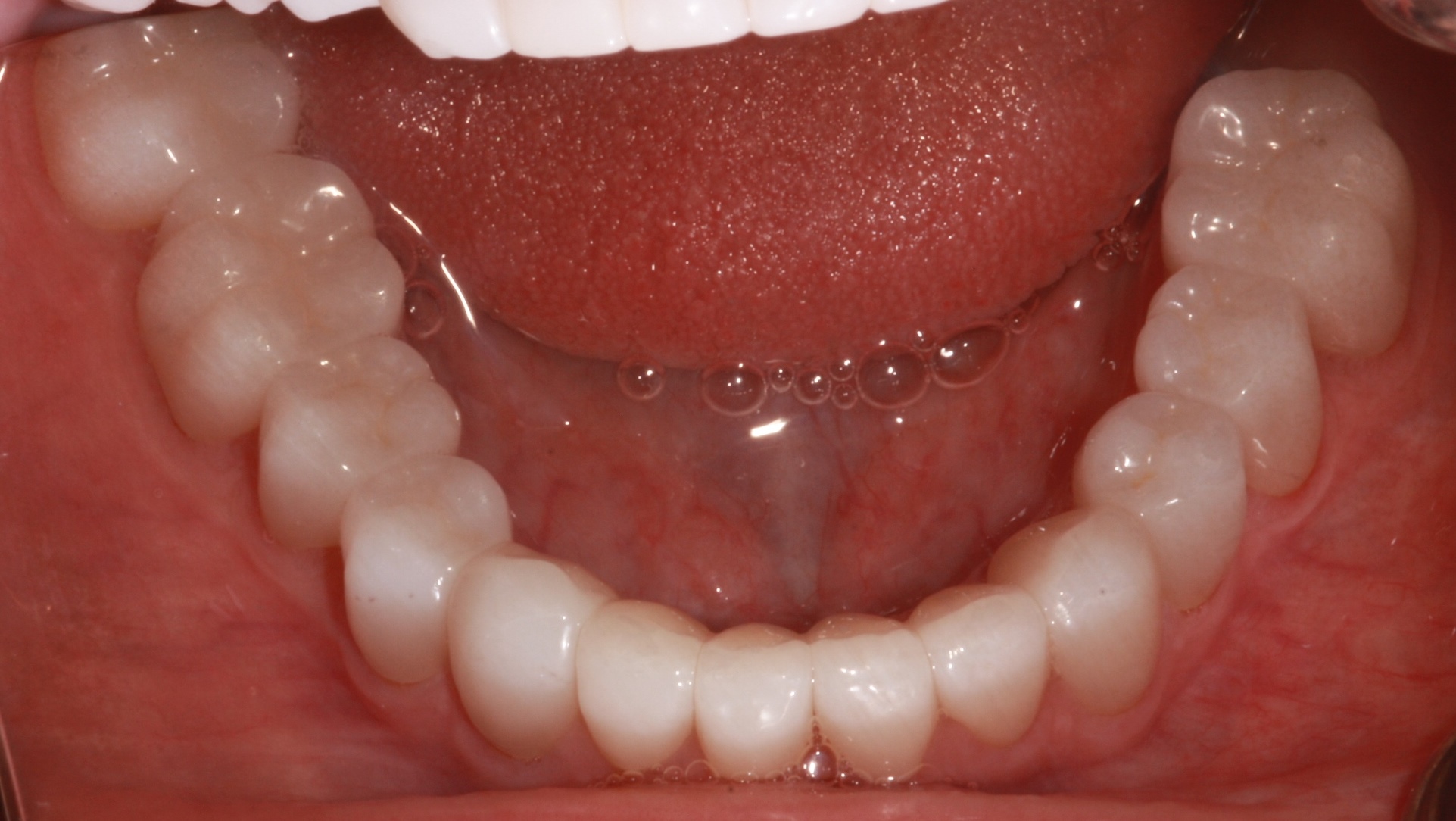
The patient also had functional concerns. She did not have a comfortable "home" position and was aware of grinding her teeth during the day. There was an extreme loss of tooth structure, and yet she was only experiencing some moderate tooth sensitivity.

During the initial interview, the patient expressed concern about the potential financial burden but seemed committed to finding a way to manage treatment.

Chief Complaints

  1. Color, shape, and size of existing teeth
  2. Prominent chin and appearing to constantly frown
  3. Moderate tooth hypersensitivity
  4. Unstable bite
Fig 1. Initial condition of the patient
Fig 2a. MIP intraoral photograph

Medical History

  1. Long-term undiagnosed gastroparesis; Now under control with meds.
  2. Celiac disease
  3. Chronic migraine headaches
  4. Insulin controlled diabetes. General difficulty in controlling glucose levels, even with meds
  5. High blood pressure
  6. Stage 3 kidney disease


Medication list:

  • Humalog
  • Tresiba
  • Altace
  • Calcitrol
  • Rayaldee
  • Levothyroxine
  • Protonix

Diagnostic Findings

A. Extra-and Intraoral Findings

i. Dental midline 2mm to the left
ii. Normal lip movement
iii. Deficient maxillary tooth display at rest, excessive mandibular tooth display.
iv. Asymmetric facial thirds with patient deficient in the lower third

B. TMJ/ functional range of motion/ muscles of mastication

i. The patient had asymmetric opening with pain bilaterally
ii. Audible joint sounds bilaterally during function
iii. Pain experienced on opening; max. open 47mm
iv. Pain in movement to the left and right
v. The patient reported jaw displacement during regurgitation episodes and in chewing. The jaw has locked at times with difficulty occluding.
vi. Acute pain on palpation of the temporalis, masseter, anterior digastric, frontalis, and strap muscles in the neck
vii. Joint pain upon loading
viii. Upon functional movements, the patient showed extreme crossover movements laterally.

C. Intraoral

i. Missing numbers 1, 16, 17, and 32
ii. Recently placed zirconia crown numbers 6 and 7
iii. Composites- number 3-DOLB, number 2-O, and number 13-O
iv. Erosion and attrition into the dentin on all unrestored teeth

Fig 2b. Noticeable two-step occlusal plane
Fig 3. Maxillary arch with signs of erosion on lingual surface
Fig 4. Mandibular arch with signs of attrition and erosion

D. Periodontal

i. Gingivitis with calculus build-up around lower mandibular anteriors numbers 23-26
ii. Gingival asymmetry due to compensatory eruption
iii. Fair plaque control and oral hygiene

Fig 5. Periodontal charting of the patient

E. Occlusal Notes

i. Pt could not repeat MIP. Lack of “home” position
ii. The first point of contact using leaf gauge as deprogrammer was a bulbous composite added to occlusal of numbers 15 and 31
iii. Group function guidance with working and non-working interferences

F. Radiographic Review (FMX from the previous dentist)

i. No periapical pathology noted
ii. no bone loss

Fig 6. Panoramic X-ray of the patient
Fig 7. Full mouth series of x-rays

G. Diagnosis and Prognosis

i. AAP Stage 1, Grade A
ii. Class II malocclusion with two-step occlusal plane, retroclined lower incisors
iii. Chronic myofacial pain, disk displacement with reduction, capsulitis
iv. Erosion with attrition
v. Day and Nocturnal Bruxism

H. Summary of concerns

Is the patient’s current dentition restorable?
How do we manage the patient’s joint disorder in conjunction with the proposed treatment?
How do we address her cosmetic concerns of a premature aging smile?
How do we manage her occlusion and VDO through treatment?
How do we help her control her diabetes?

Proposed Treatment

FULL MOUTH CROWN AND BRIDGE RECONSTRUCTION WITH FLAPLESS CROWN LENGTHENING AND OPENING OF VERTICAL DIMENSION

  • Phase I- Formulation of proposed restorative design using digital iOS scanning.
  • Phase II- Delivery of a functional bonded mock-up to test aesthetic design and provide therapeutic treatment for the joint while establishing consistent MIP.
  • Phase III- Evaluation of occlusion and joint stability in functional provisionals with occlusal adjustment prior to preparation.
  • Phase IV-iOS capture of adjusted provisional with updated design that meets the patient’s agreed-upon aesthetic goals.
  • Phase V- Preparation of all teeth using functional mock-ups as preparation guide.
    • Gingivectomy to alter gingival margins on numbers 7 and 8
    • Scan for final impressions.
    • Delivery of temps from previously designed mock- up
  • Phase VI-Monitor at 8 weeks for gingival healing. Modify and rescan preparations for numbers 7 and 8, if margins become exposed.
  • Phase VII-Fabrication of definitive zirconia restorations and delivery.
  • Phase VIII- Splint therapy with adjustments at 6-month intervals in addition to periodontal recare.

Active Clinical Treatment

Phase I

The patient’s full arch was scanned with 3SHAPE iOS, and placed in a leaf gauge, which was opened to 8 mm to simulate the desired amount of VDO increase in the anterior segments. This would allow approx. 4 mm of increase posteriorly, allowing for additive mock-up and minimally planned preparations. Under the guidance of an assistant, the patient went through protrusive and retrusive movements for 30 minutes without any tooth-tooth contact. This was done to deprogram the muscles and to seat the condyles at the proposed increased VDO. A bite was then taken at the proposed vertical, in retrusive position, with the leaf gauge in place using iOS for the creation of the digital design.

The digital design was fabricated to lengthen the central incisors to 10.5 mm, and the gingival margin was raised 2.5 mm on number 8 and 2 mm on number 7. The midline was shifted 2 mm to the right in the maxillary arch. Posterior segments were raised in the mandibular arch to level the occlusal plane and to decrease the stepped occlusal plane in the mandible. The gingival margins of the mandibular incisors were not altered despite compensatory eruption, as they were not in the aesthetic zone.

Phase II

Putty Stents, scalloped to the proposed gingival heights, were fabricated from 3-D models based on the digital design. Delivery of the functional bonded mock-up was completed using the stents for transfer. Teeth were conditioned with universal self-etching adhesive material prior to delivery to aid in retention on the facial and lingual. The bulbous composite on number 15 was reduced prior to the placement of the mock-up. A bis-acryl mock-up in the proposed shade B-1 was adjusted after placement with the leaf gauge to remove working and non-working interferences and to ensure bilateral posterior contact in MIP, with anterior guidance in excursive movements. Special consideration was given to the lateral incisors, which had to be reduced in extreme crossover from the original design.

Fig 8. Intraoral Mock-up before preparations

Phase III

The patient was seen at weekly intervals for occlusal adjustment, altering the shape of the teeth until functional movements were balanced in lateral and protrusive excursions. The initial design was too steep, and the anterior guidance had to be adjusted to accommodate the functional movements of the patient. The canines were adjusted in length until crossovers were managed on the lateral and central incisors. The patient reported significantly less pain in joints within 5 days after placement of mock-up. Canines initially showed extensive wear with hypersensitivity. Guidance was decreased by minimizing the angle of disclusion in protrusive and anterior-lateral excursions. This was adjusted in small increments over 6 weeks until the patient was comfortable. Pt reported a major reduction of headaches at week 5, with the elimination of pain on opening and working movements at week 6. Pt was delighted with aesthetics and improved masticatory function. New iOS scans were made at week 6 once the patient was pleased with the outcomes and the TMJ was comfortable. This captured the changes in the functional bonded mock-up and served as the new design template moving forward.


Phase IV: Prep and Surgical Intervention

Pt was pretreated with muscle relaxer, cyclobenzaprine prior to the appointment. Continuous glucose monitoring was used during the appointment. Preparations were made through the mock-up using reduction wheels and slots to facilitate rapid reduction while only reducing necessary tooth structure. After grooves were made, the functional bonded mock-up was removed, and the preparations finalized. All teeth were prepared for full coverage restorations, though minimal reduction was needed due to the nature of increased VDO and addition-based design.

  • Maxillary and Mandibular Posterior segment reduction was accomplished first. The mock-up served as a preparation guide.
    Reduction slices were made through the temporary material. The temporary material was then removed, interproximal slices were made, and reduction lines were connected in all planes.
Fig 9. Mock-up used as a preparation guide to allow for more conservative preparations
  • A Diode laser was used to reduce 2.5 mm of gingival tissue on number 8 and 2 mm on number 7. Due to invasion of biological width, a wedelstaedt chisel was used to reduce 1.5 mm of facial bone without raising a flap. Hemostasis was obtained with pressure for 5 minutes.
  • A full-arch scan was made of the posterior maxillary and mandibular preparations. A bite was captured at the maintained VDO since anterior segments were not yet prepared from the mock-up. After scans were completed and put in 3-D relationship with each other via iOS, the anterior segment of scans was deleted, anterior teeth prepared, and then re-scanned.
Fig 10. Posterior teeth prepared with anterior mock-up in place to maintain proposed VDO and allow for scanning with the ideal VDO
Fig 11a. Full upper arch preparations
  • The teeth were then temporized, and the occlusal scheme was confirmed.
Fig 11b. Full mouth provisional restorations

Phase V: Post-operative care

Gingival healing was monitored for 7 weeks. Tooth margins did not become exposed post-operatively. A stable occlusion was confirmed, and the joints were comfortable. We then proceed to the final restorative phase of treatment.

Phase VI: delivery of final restorations.

All restorations were fabricated with Sagemax Monolithic Zirconia-Shade BL-4. Intaglio of restorations were conditioned with Z-Bond Zirconia Primer. Restorations were all delivered with Ivoclar Speedcam self-adhesive resin cement with a standard sextant approach. The lingual incline of number 6 had to be adjusted slightly to the patient’s comfort. No other adjustment was required. The digital design allowed for the contours of the functional design we achieved in the mock-up to be replicated very accurately.

Fig 12a. Maxillary arch with final restorations
Fig 12b. Mandibular arch with final restorations
Fig 12c. Frontal view of the full-mouth reconstruction with a re-stablished CDO

Commentary

Initial documentation, in this case, was not as complete as what would ideally be collected. Although adequate, the FMX was not as diagnostic because the patient’s TMD limited her ability to position her teeth appropriately. This did not alter the final plan.

There are admittedly different ways that treatment could have been sequenced. The beauty of the functional bonded mock-up is that segments could be managed independently from each other to facilitate phased treatment, if necessary.

Having just begun my own journey into 3-D printing, I was hesitant to utilize the technology. Now, with more experience, I would have considered printing the temporaries in small segments coordinating with my laboratory to facilitate cleanings during an extended period of treatment.

An unanticipated additional benefit of the mock-up was that it instilled confidence in the patient to proceed with treatment. Her previous dentist had convinced her that her dentition was not salvageable. In more difficult and complex case situations, motivating and getting the patient excited about treatment is often essential for final success.

The finances were not easy for this patient. Although costly, this treatment approach became far less of a burden than what would have been required for an implant-supported reconstruction. She was very pleased with the result. The total treatment time was approximately 3 months.

Fig 13a Final smile photograph
Fig 13b. Lateral view which denotes harmonious relationship between the maxillary and mandibular arch