Case 12: Special Report 12

We are publishing this special report because of the interesting way the maxillary arch was digitally planned and efficiently treated. The mandibular arch received only treatment necessary to create an ideal occlusal plane. Future mandibular arch restorative treatment is planned.

Digitally Designed and Fabricated Teeth in a Day

A 63-year-old female patient visited the private clinic in Overijse, Belgium. The patient was healthy, was under no medication, and quit smoking seven years ago. The patient’s chief complaint was marked increased mobility of the upper teeth and inferior esthetics. The patient wished for an esthetic and preferably fixed solution that would enable her to eat, interact and maintain hygiene properly.

Data Acquisition

  • Medical and dental history.
  • Comprehensive clinical (extra-oral, intraoral, dental, periodontal, functional, esthetic) and radiologic examinations were performed. Standardized extra-oral and intraoral photographs of the initial situation were taken.
  • Intraoral scans of both jaws (3Shape, Copenhagen, Denmark).
  • Panoramic radiographs were performed.
  • Facilitated communication with treatment team. All data was uploaded to a cloud-based platform (SmileCloud, Dentcof, ADN3D Biotech srl, Timisoara, Romania).
Initial full face
Initial view in maximum intercuspation
Initial maxillary arch
Mandibular arch

Diagnosis

Based on the clinical and radiographic findings, the following diagnoses were determined.

  • Extraoral: Non esthetically-pleasing smile, no facial asymmetries.
  • Intraoral: No abnormalities.
  • Dental: Multiple insufficient restorations, multiple secondary and cervical caries.
  • Periodontal: Terminal Stage IV Grade A Periodontitis.
  • Function: Sufficient contacts in maximal intercuspation position, canine-protected dynamic guidance.
  • Radiographic: Generalized horizontal bone resorption, multiple insufficient restorations and root canal treatments, multiple secondary caries and multiple periapical lesions.
  • Prosthetic: Insufficiently restored adult dentition.
  • Esthetic: Compromised esthetics.

Based on the diagnosis, the prognosis of the upper teeth was set as poor, leading to a case of terminal dentition.

The available maxillary treatment options; complete denture, implant-retained removable dental prosthesis, implant-supported removable dental prosthesis, implant-supported fixed partial denture were presented to the patient along with their advantages, limitations, required treatment time, as well as financial requirements. In addition, the initial plan of the smile design was presented to the patient to educate her about the different possibilities of tooth shapes, as well as proposed smile outcome. The patient chose the option of implant-supported fixed partial denture.

Active Clinical Treatment

Hygiene Phase

Education and instruction of the patient to optimize her oral hygiene.

Pre-Prosthetic Phase

The initial extraoral and intraoral photographs were matched using cloud-based algorithms and helped to provide an initial smile design with individual tooth shapes (SmileCloud, Dentcof, ADN3D Biotech srl, Timisoara, Romania).

A cone-beam computed tomography was performed (Pax i3D- Vatech, Seoul, South Korea). The CBCT data was imported into an implant planning software, along with intraoral scan data. In addition, STL data of the future restoration was uploaded and matched in an implant planning software (MSOFT by SMOP, MIS Implants, -Bar Lev, Israel). Afterwards, the data was used to virtually plan implant positions in the upper jaw.

SmileCloud design with natural shapes

The teeth were virtually removed except for nos. 5, 7, 10 and 13. The remaining teeth were kept to serve as temporary stabilizers of the surgical guide during the immediate implant placement procedure. Then, seven implants were virtually planned in the maxilla in positions of nos. 4, 6, 8, 9, 11, 12 and 14. Multi-unit abutments (MU, MIS, Bar Lev, Israel) were virtually connected to the implants nos. 4, 6, 11, 12 and 14 and CONNECT abutments (CONNECT, MIS, Bar Lev, Israel) were virtually connected to the implants nos. 8 and 9. In addition, four anchor pins were virtually planned for the implant surgical guide, which will serve in positioning of the immediate temporary restoration at a later time point. The pins were distributed in between the implants in the palatal area.

The planning procedure and manufacturing of the surgical guide as well as temporary FPD were completed.

Surgical Phase

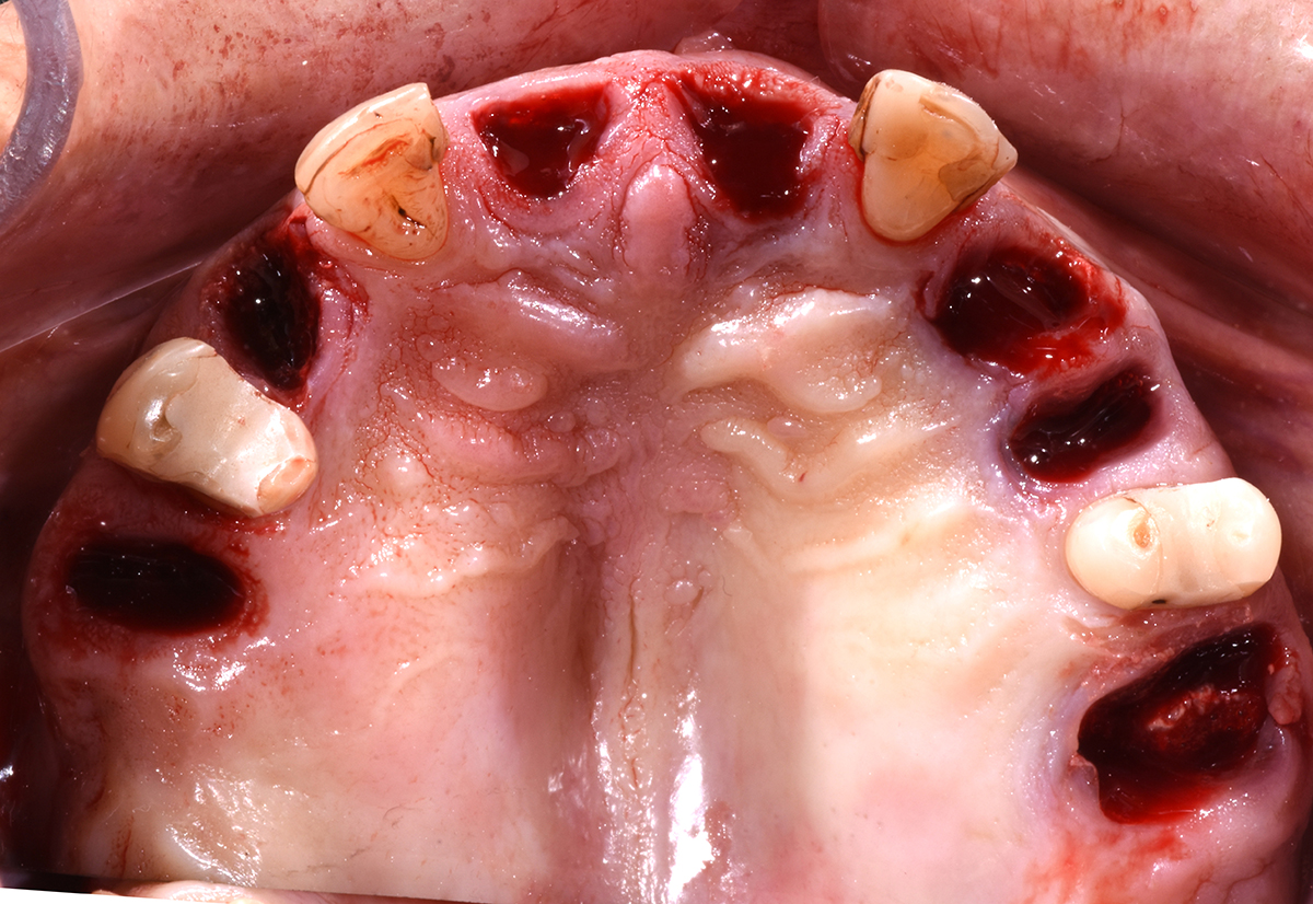
Extraction of teeth nos. 4, 6, 8, 9, 11, 12 and 14 was completed.

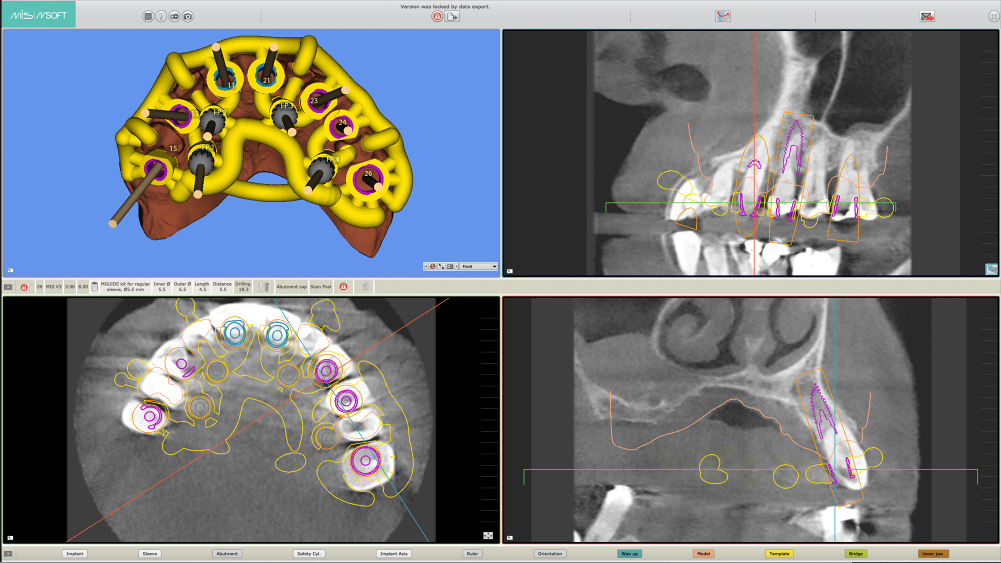
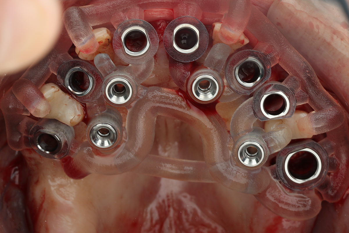
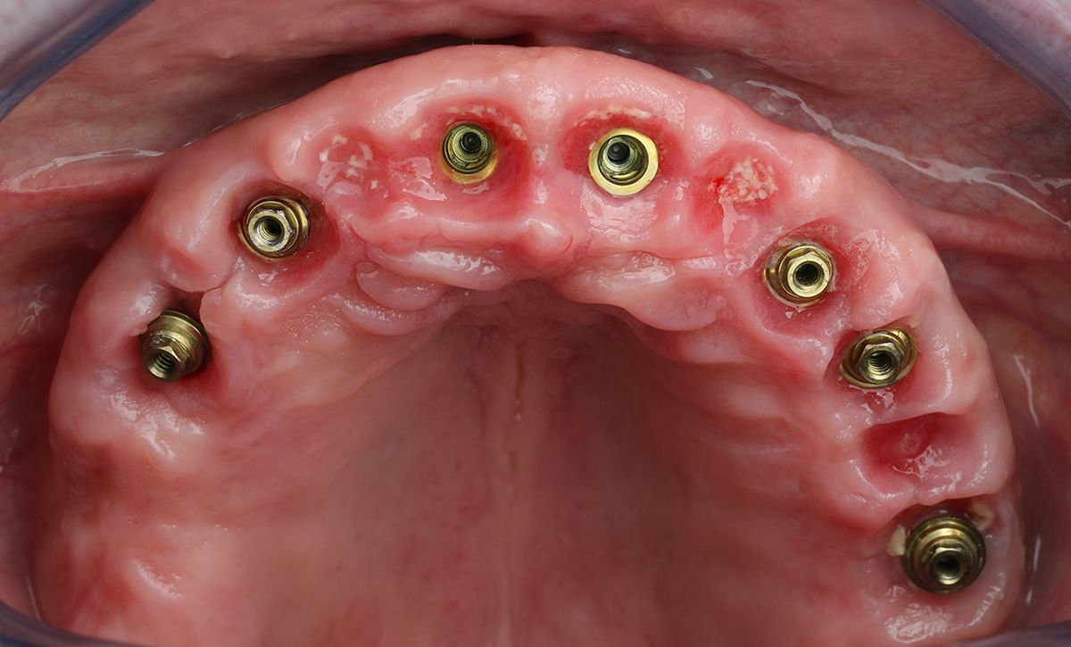
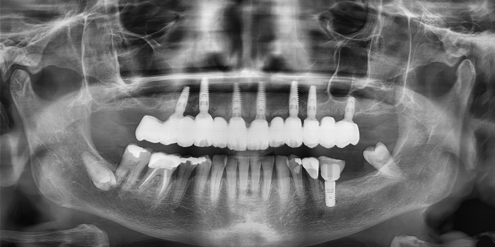
The implant surgical guide was secured onto the remaining teeth and the fit was verified. Afterward, implant site osteotomies were created using sequential drills, followed by fully guided implant placement (MGUIDE, MIS Implants, Bar Lev, Israel) protocol. In total, seven implants were placed in the maxilla. (V3, MIS Implants, Bar Lev, Israel)

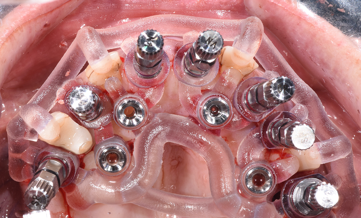
After all implants were installed, drilling of the anchor pins took place. Then the surgical guide was removed and the remaining teeth nos. 5, 7, 10 and 13 were extracted.

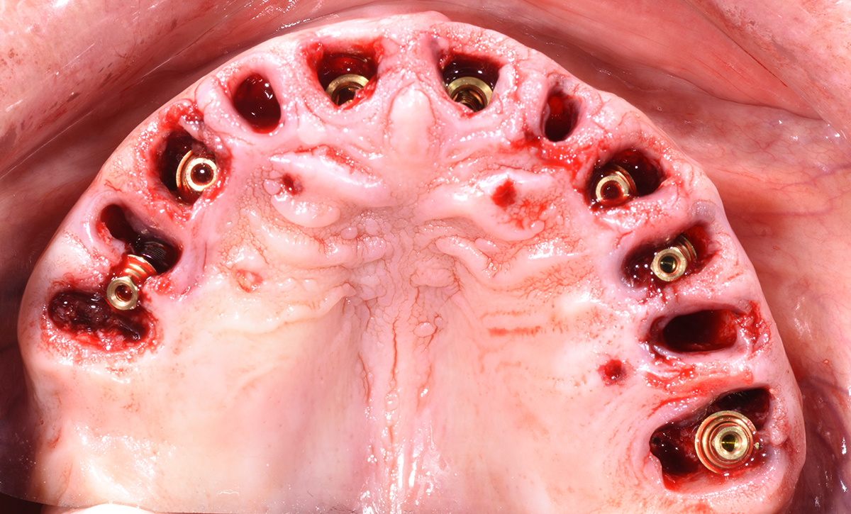
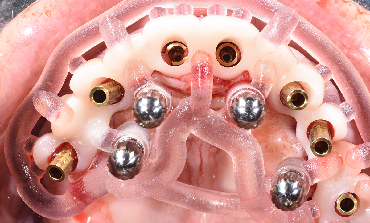
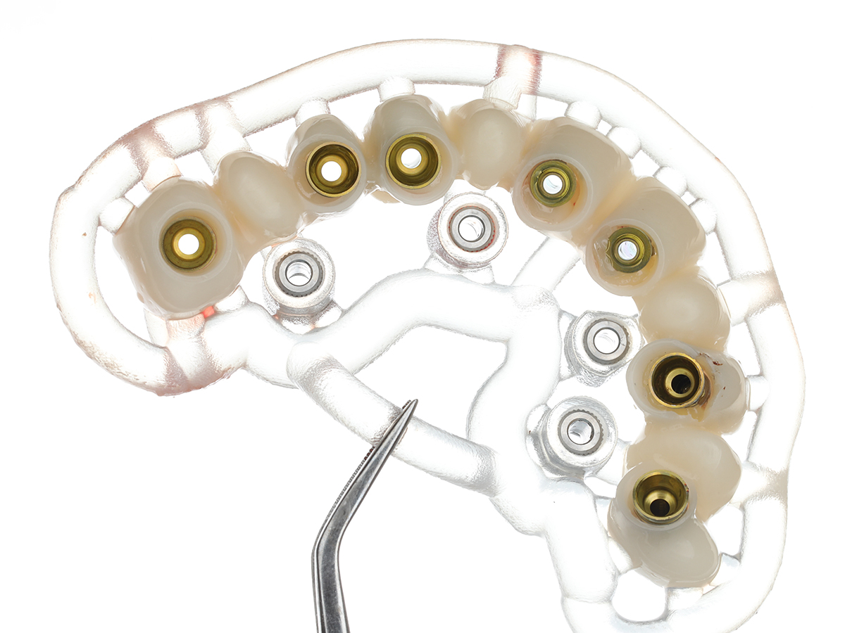
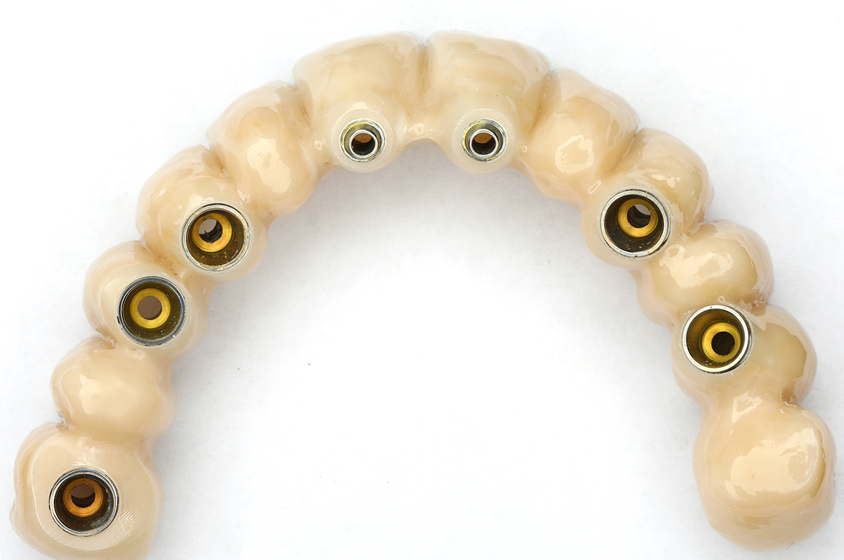
The primary stability of all implants exceeded 45 Ncm. Multi-unit and CONNECT abutments (MIS Implants, Bar Lev, Israel) were attached to the implants according the pre-prosthetic plan and were torqued to 35Ncm. Afterward, the temporary FPD was loaded into the positioning appliance and secured in the planned position using the anchor pins.

Selective extractions
Surgical stent positioned by remaining teeth
Implants placed with surgical stent
Anchor pin locations

The temporary cylinders were screwed onto the multi-unit abutments and the voids with the access holes in the temporary FPD were filled using a flowable composite (Filtek Z250XT, 3M, St Paul, USA) in the upper third of the access holes.

After all cylinders were attached, the anchor pins were removed, followed by the positioning appliance and the FPD. The remaining voids between the cylinders and the temporary FPD were filled extraorally with PMMA resin (Unifast, GC, Tokyo, Japan).

After final contouring was completed the temporary FPD was screwed into place, the occlusion was fine tuned, and post-operative instructions were given, including having soft diet for eight weeks after the procedure.

Provisional and positioning stent oriented with anchor pins
Temporary cylinders luted to provisional
Post surgical maximum intercuspation
Twelve weeks after surgery

After 12 weeks of healing, conventional impressions using impression copings and a polyether material (Impregum, 3M Espe, Seefeld, Germany) along with custom-made impression trays were made.

STL files were imported into the design software (exocad, exocad GmbH, Darmstadt, Germany) and superimposed with the design files in order to construct the final restoration. After the design was finalized, the final FPD was milled out of a zirconia material (Prettau, ZirkonZahn ,Gais, Italy). Then the FPD was individually colored and sintered to final dimensions. Further esthetic enhancements were carried out by applying veneering ceramic to the facial surfaces of all teeth.

After verification of the function, esthetics, phonetics and hygiene, the final FPD was delivered onto the implants. Two weeks after delivery, the patient was recalled for verification of the integrity of the restorations. The patient reported no issues with the newly delivered restorations and is very satisfied with the outcome.

Final prosthesis
Definitive maximum intercuspation

Conclusion

Definitve full face

This case report presents a workflow that aims to improve the accuracy of immediate guided implant surgery and simplifies the procedure of delivering immediate temporary restorations for edentulous arches. The simplification of the clinical procedures and the immediate approach shorten the treatment time, reduce the patient’s morbidity, and improve the overall quality of life. Although the clinical procedures can be significantly simplified, the digital workflow presented requires an experienced and well-trained treatment team.

Acknowledgments

The authors would like to thank MIS Implants MCENTER (MIS Implants, Bar Lev, Israel) for their support in the design and manufacturing of the surgical guide, positioning appliance, and immediate temporary FPD, as well as  the support of MetaLab (Timisoara, Romania) for the design and manufacturing of the final rehabilitation.

Dr David Norré is both a prosthodontist (University of Leuven) and specialist in implant dentistry (University of Liège), practicing in Overijse, Belgium. He is a visiting faculty member the University of Leuven.