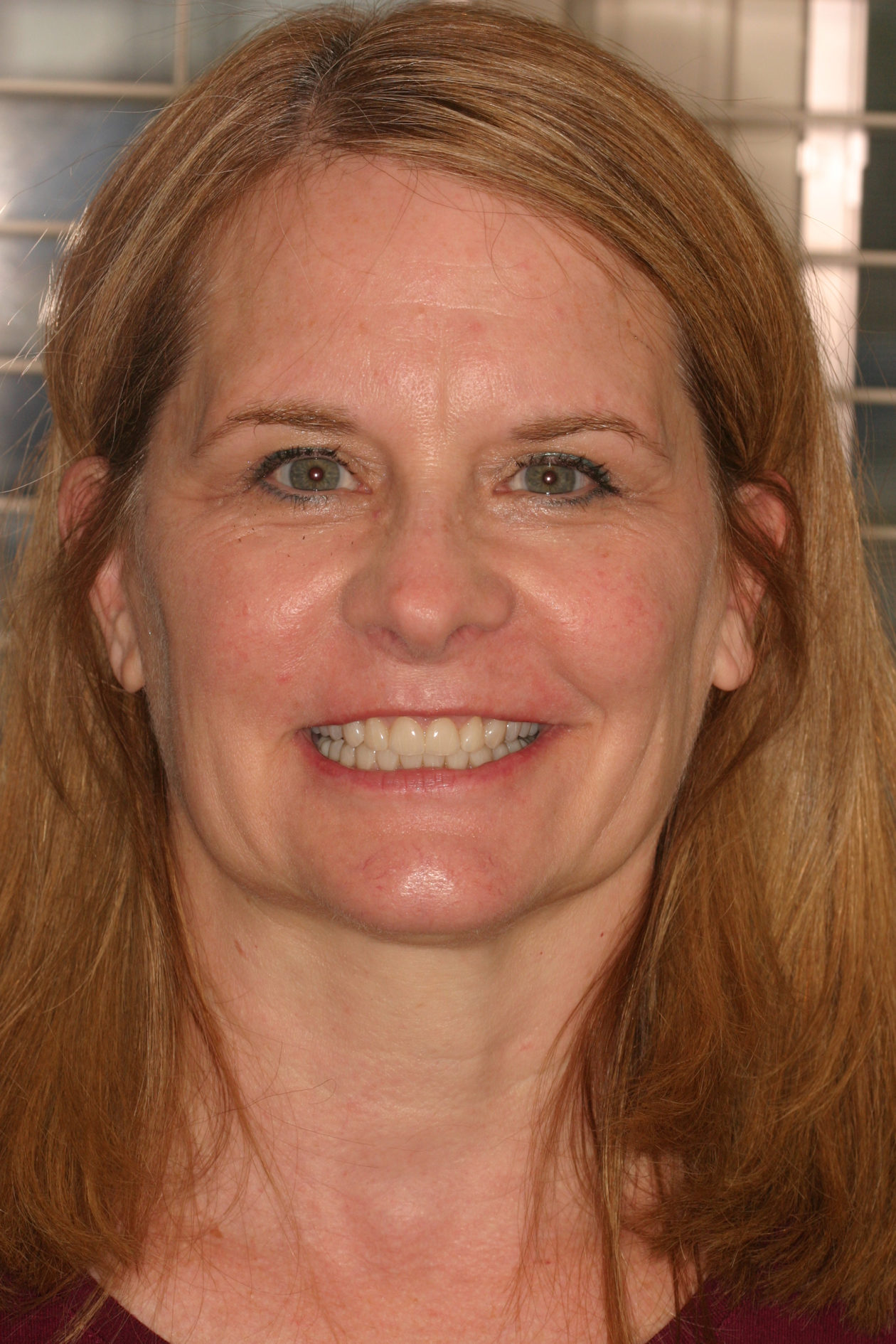
Case 99: Restoration of a Worn Dentition Without Orthodontics

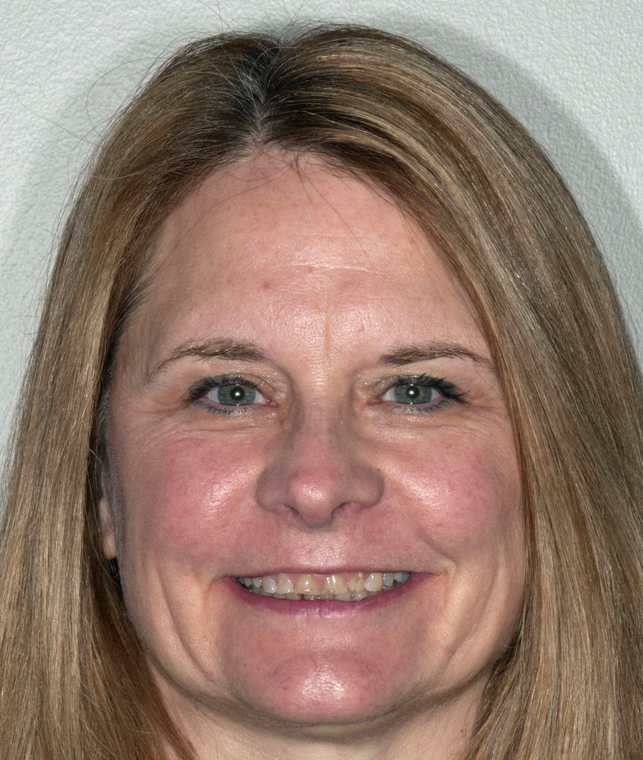
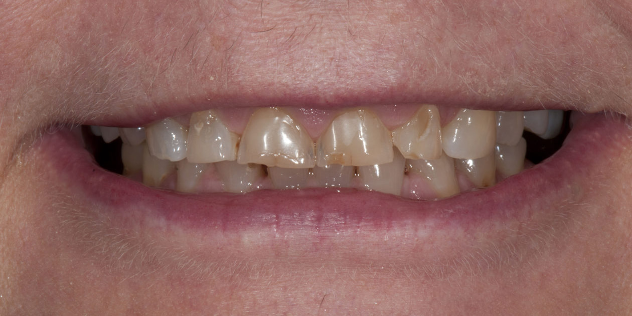
The patient was self-referred to the periodontist’s office. She had not sought dental care for more than 10 years. The patient’s chief concerns included: 1) “My front teeth are chipping and wearing down.”; 2) “I do not want to smile anymore.”; and, 3) intermittent pain associated with tooth no. 6.

After initial treatment at the periodontist’s office, the patient was referred for an endodontic evaluation of tooth no. 6. The endodontic treatment was delayed but ultimately completed nine months later (March 2017). The patient did not return to continue her dental care for an additional seventeen months (August 2018). The patient had become engaged to be married and was highly motivated to proceed with comprehensive care.

Medical History

  • Medical history is non-contributory.
  • The patient has seen her gynecologist, but not a family physician for more than five years.
  • The patient denies taking over-the-counter or prescription medications.
  • The patient expresses extreme anxiety toward dental care.

Diagnostic Findings

Extraoral/Facial

  • Facial symmetry within normal limits.
  • Facial vertical proportions are balanced: Middle third: 54mm; Lower third: 58mm.
  • Maxillary dental midline is coincident with facial midline.
  • Nasolabial angle is obtuse.
  • Upper and lower lip prominences are favorable to esthetic plane.

Smile Analysis

  • Maxillary central incisor display at rest: -2mm.
  • Gingival display upon maximum smile: 3mm.
  • Upper lip length: 20mm.
  • Upper lip mobility: 10mm.
  • Smile arc is coordinated with asymmetric lower lip.

TMJ/Mandibular Range of Motion/Muscles of Mastication

  • Right TMJ: clicking with disc displacement.
  • Range of motion within normal limits.
  • No muscle tenderness detected.
  • No history of TMJ pain.
  • Dental wear patterns: protrusive and eccentric facets present.
  • Patient experiences trismus with extended mouth opening.
  • Requires bite block during dental treatment for mandibular support.

Intraoral Findings

Dental

  • Maxillary incisor dimensions: 6.5mm length, 8mm width.
  • Triangular coronal anatomy of maxillary anterior teeth.
  • Tooth no. 10: rotated 15-20 degrees.
  • Diastema: teeth nos. 25-26.
  • Missing teeth nos. 1, 2, 16, and 30.
  • Impacted teeth nos. 17 and 32.
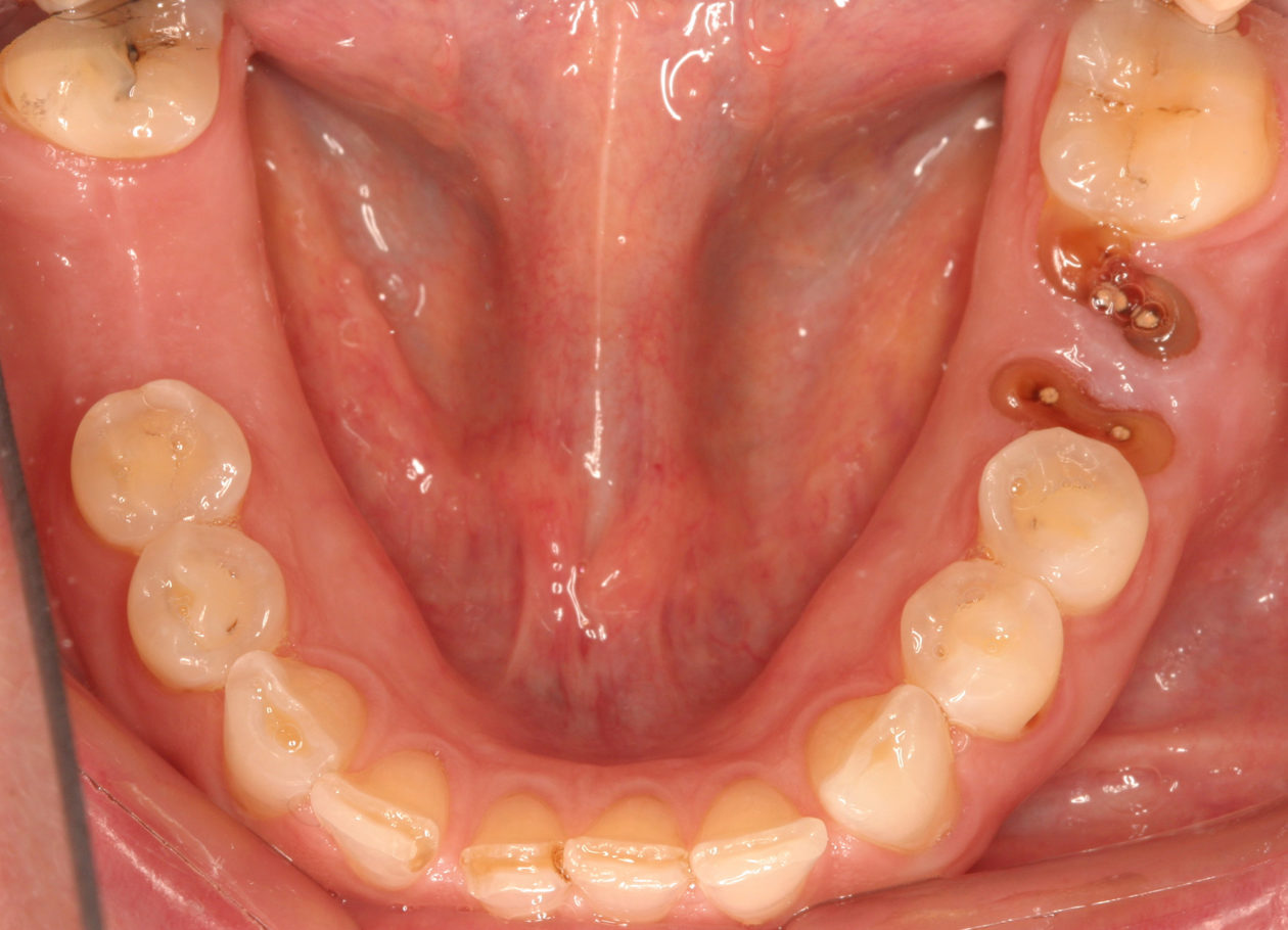
  • Severely decayed tooth no. 19 with residual root fragments.
  • Multiple areas with cervical decay.
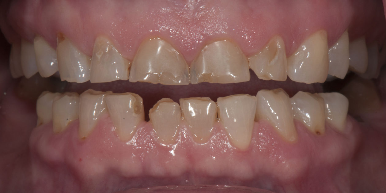
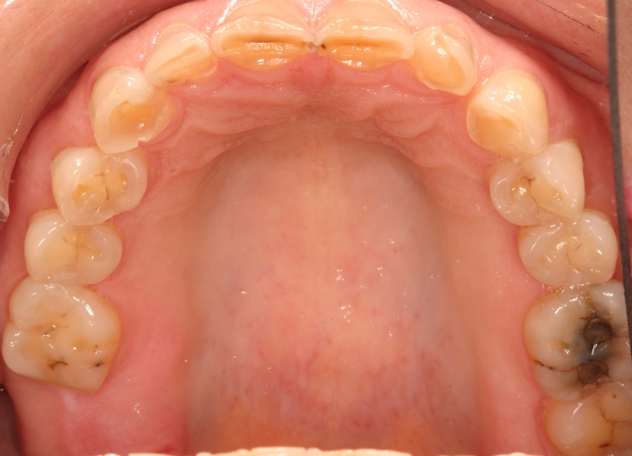
  • Evidence of advance attrition and erosive tooth structure loss.
  • Maxillary/mandibular anterior/posterior wear has resulted in short clinical crown lengths.
  • Chipped and fractured maxillary and mandibular incisors.

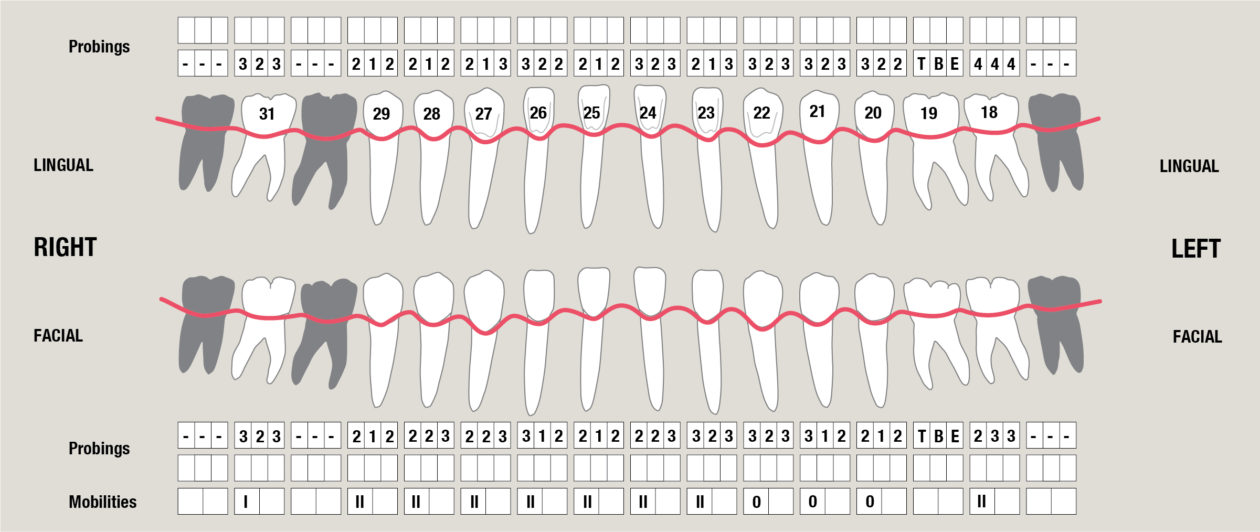
Periodontal

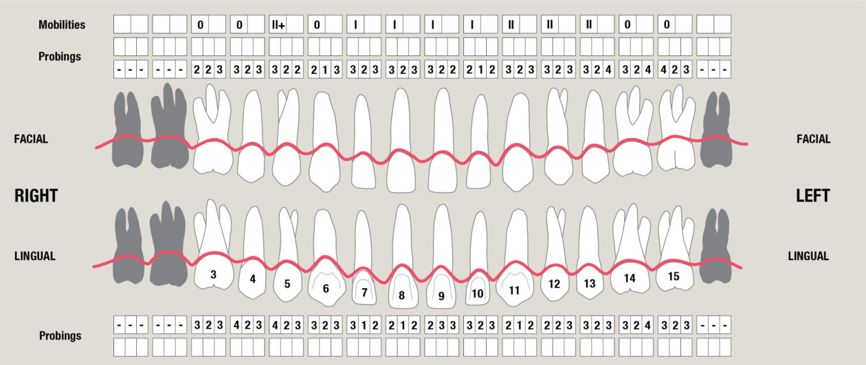
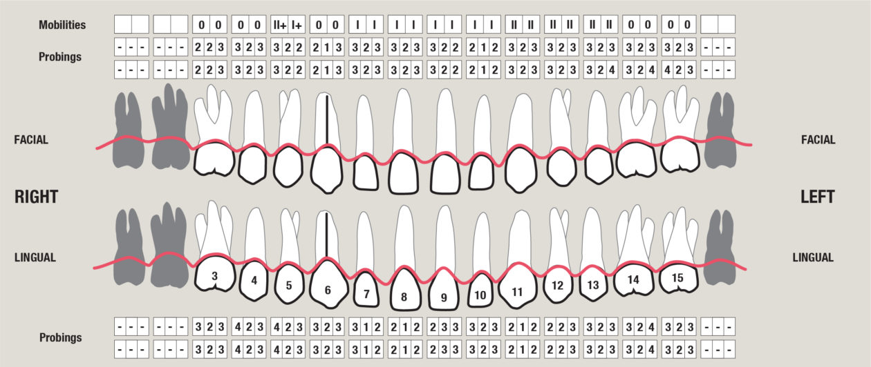
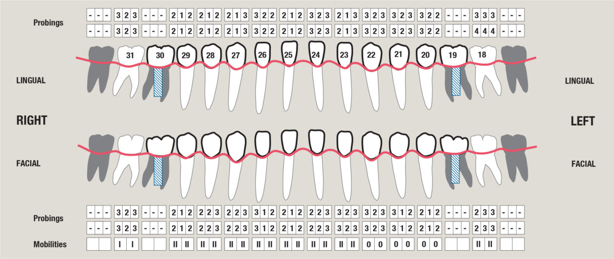
  • AAP classification: stage I, grade A.
  • Clinical attachment levels are stable.
  • Probing depths all 4mm or less.
  • No gingival recession present.
  • Mobility: generalized Cl II mobility.
  • Thick gingival phenotype.
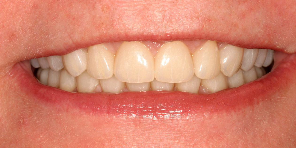
Initial smile.
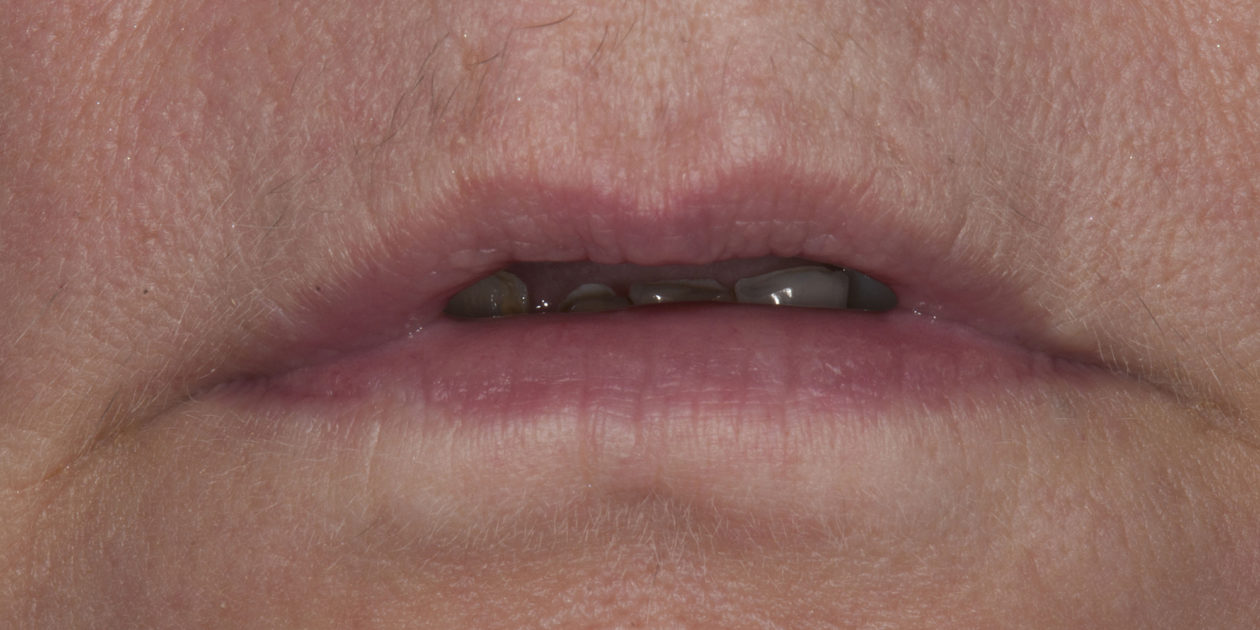
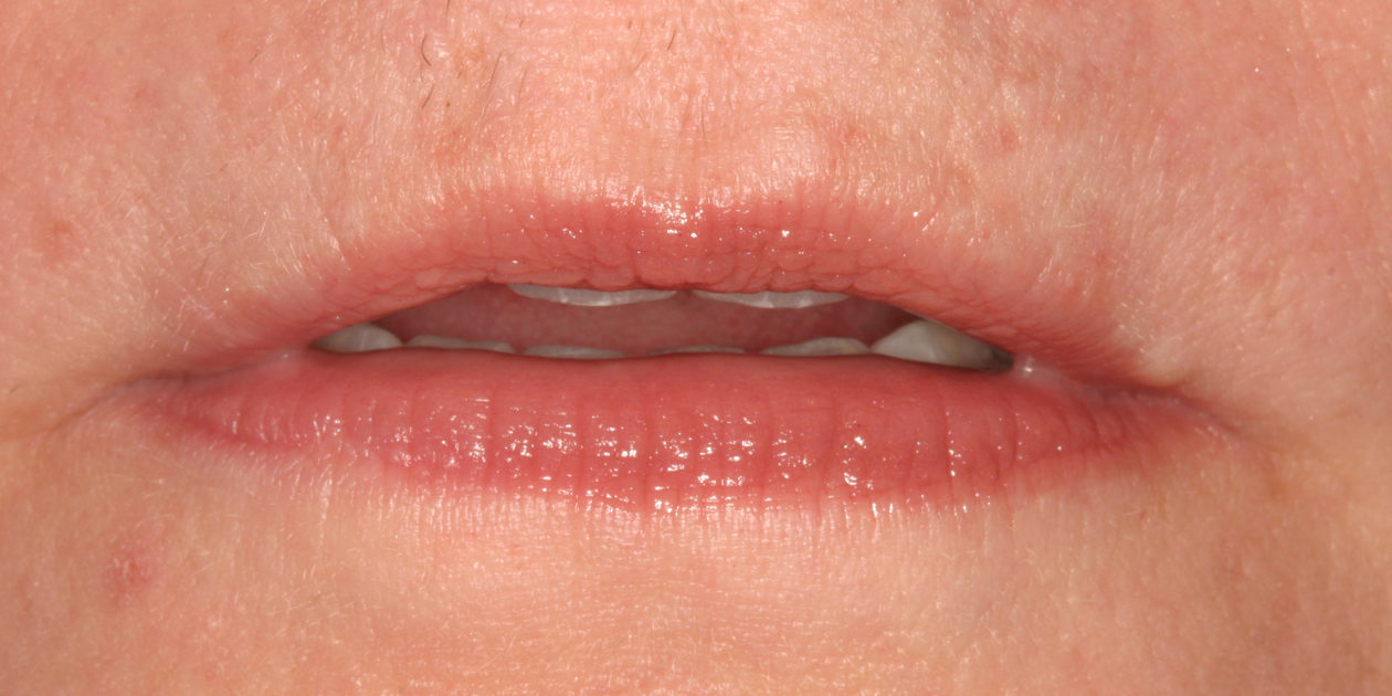
Initial tooth display at rest.
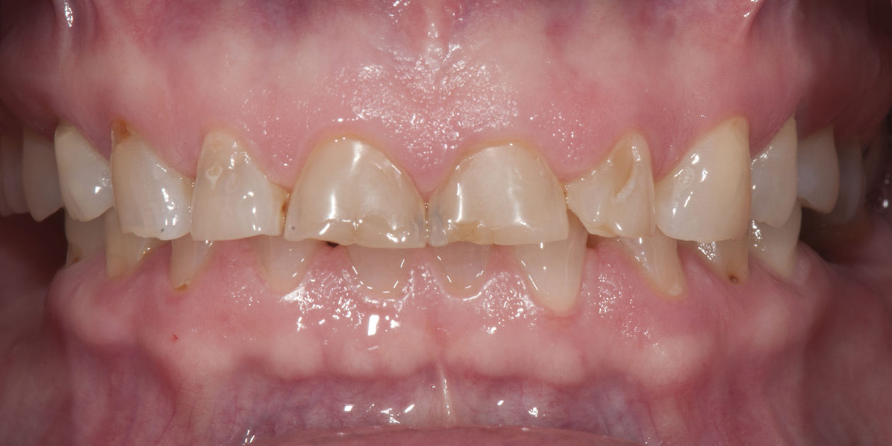
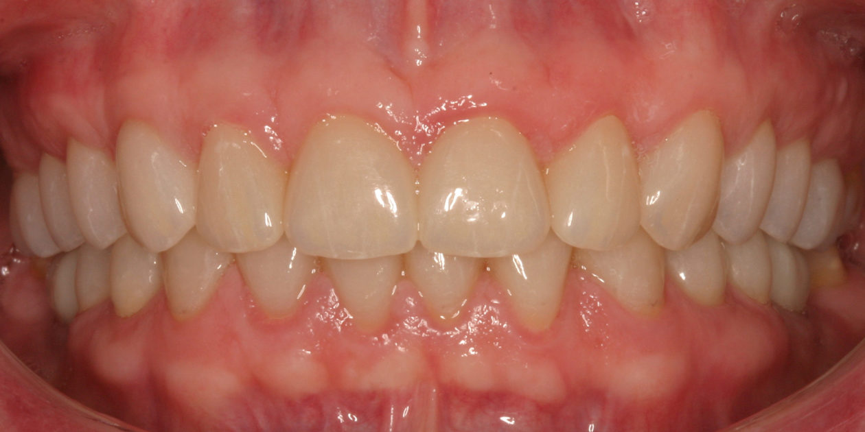
Initial maximum intercuspation.
Initial occlusal planes.
Initial maxillary arch.
Initial mandibular arch.

Occlusal Notes

  • Centric premature contact: teeth nos. 3/31.
  • CR/CO slide: 4mm anterior.
  • Angle’s Cl I canines.
  • Generalized Cl II mobility due to primary occlusal trauma.
  • Supra-eruption tooth no. 3.
  • Moderate dento-alveolar extrusion teeth nos. 7-10.
  • Generalized attrition and erosion.
  • Possible loss of vertical dimension.
  • Multiple anterior diastemas.

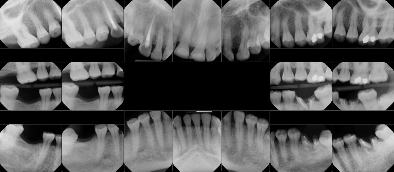
Pre-Operative FMX

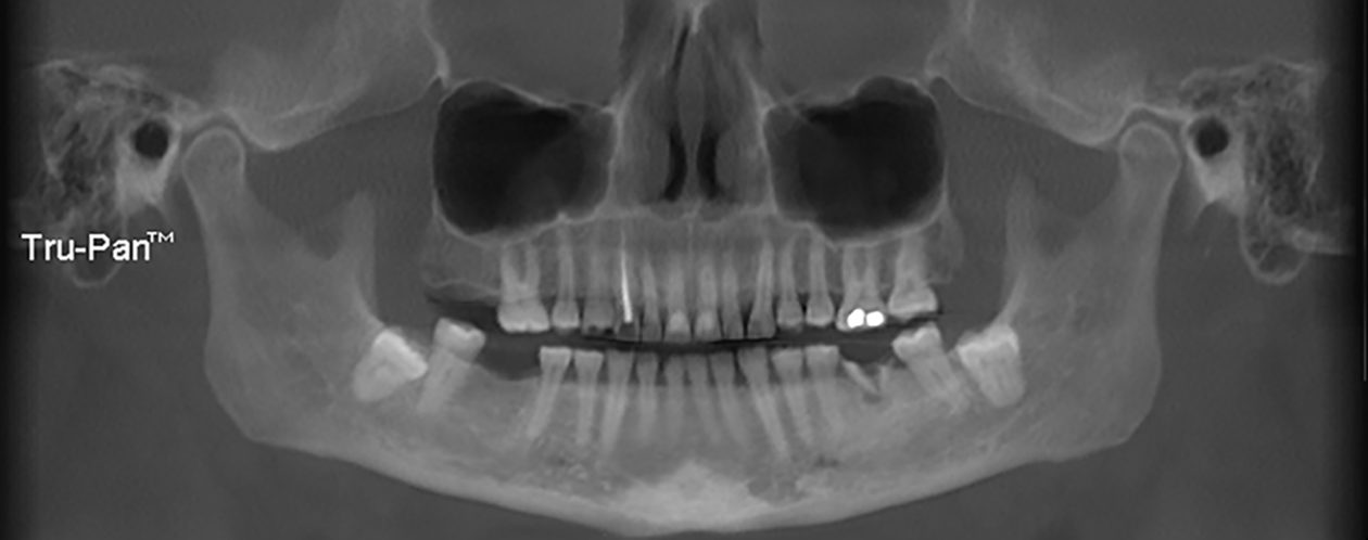
Pre-Operative 3D Scan Panoramic X-ray

Radiographic Review

  • Alveolar bone heights: within normal limits.
  • Full bony impactions teeth nos. 17 and 32.
  • Root anatomy: generalized tapered and moderately short roots.
  • Crown anatomy: triangular maxillary/mandibular incisors.
  • Fractured and decayed root fragments with associated periapical lesion: tooth no. 19.
  • Edentulous areas of teeth nos. 2 and 30.
  • Advanced wear: anterior teeth and premolars.
  • Decay in anterior areas.
  • Pulpal exposure tooth no. 6.

Diagnosis and Prognosis

Diagnosis

  • Localized mild gingivitis: Dental biofilm-induced AAP Stage I; Grade A.
  • Unidentified acidic accelerant influencing erosion.
  • Protrusive and eccentric bruxism.
  • Loss of vertical dimension.
  • Loss of maxillary incisor display at rest and smiling.
  • Compromised esthetics.
  • Dental phobia.

Prognosis

  • Periodontal: excellent.
  • Biomechanical/structural: good.
  • Functional: fair.
  • Esthetic: excellent.
  • Biologic: guarded.

Summary of Concerns

  • Is orthodontic treatment necessary to achieve an acceptable esthetic restorative outcome?
  • Can an esthetic restorative outcome be achieved given the presenting tooth morphologies, interdental spaces, and tooth sizes?
  • In order to preserve the restorative outcome, what will be necessary to manage the patient’s parafunctional habits and history of erosion?
  • Will the patient be able to tolerate an increased vertical dimension given her existing TMJ findings and present oral habits?
  • Will the patient be able to comply with long-term maintenance and occlusal therapy (bruxism appliance) given her dental phobia?
  • Can the unknown acidic component of erosion be effectively managed by restorative care?

Proposed Treatment Plan

Phase I: Diagnostic Workup

  • Obtain digital diagnostic casts, centric relation records, clinical and periodontal charting, and intraoral and CBCT radiographs and photographs.
  • Complete a combined interdisciplinary treatment plan consult with periodontist and prosthodontist.

Phase II: Esthetic Analysis

  • Establish diagnostic wax-up on mounted casts in centric relation.
  • Present to patient for outcome simulations and motivation. Receive approval.
  • Fabricate surgical stent to facilitate pre-prosthetic periodontal surgery.

 Phase III: Periodontal Therapy

  • Establish periodontal health and idealize plaque control habits.
  • Periodontal esthetic and functional pre-prosthetic crown lengthening to facilitate prosthetic goals.
  • Extraction of tooth no. 19 with guided bone regeneration (GBR).
  • Implant placement in areas of nos. 19 and 30.
  • Note: endodontic therapy was completed on tooth no. 6 eighteen months prior to restorative treatment.

Phase IV: Treatment Presentation

  • Presentation of full-mouth rehabilitation including implant placement in teeth nos. 19 and 30. To improve the esthetics and create restorative room, the vertical dimension will be opened along the centric relation hinge axis.

Phase V: Definitive Prosthetic Treatment

  • Restore all existing teeth with full coverage restorations at a newly established vertical dimension.
  • Restore implants nos. 19 and 30.

Phase VI: Maintenance

  • Provide 3-4 month dental prophylaxis re-care.
  • Bruxism appliance.
  • Compliance monitoring.
  • Erosion monitoring and treatment as indicated.

Active Clinical Treatment

Review of Treatment Goals

The patient had not seen a dentist for more than 10 years due to fear that she would lose her teeth. She was self-conscious and embarrassed of her smile. She was very concerned with the progressive wear on her teeth. She wanted to improve her smile and function before her planned wedding in the fall of 2019. The patient was referred to the prosthodontist by the periodontist.

Active Clinical Treatment

Phase I: Treatment Presentation

  • We discussed with the patient the extensive nature of her treatment that would involve restoring all of her teeth, not just the anterior teeth.
  • This would reestablish her smile, while at the same time restoring the occlusion close to her original position or vertical dimension that had been lost due to extreme wear.
  • We discussed the facial esthetic impact that reestablishing vertical dimension would provide.
  • We discussed how we are addressing and mitigating the causes of her present dental condition with our treatment design and maintenance therapy.

Phase II: Diagnostic Workup

  • Consultation with the periodontist, prosthodontist and ceramists.
  • Full-arch maxillary and mandibular intraoral impression scans and bite registration obtained (Trios, 3Shape).
  • Complete series of photographs and radiographs obtained.
  • Team of ceramists designed a digital diagnostic wax-up.
  • The ceramist provided a putty matrix based on the digital design for a motivational mock-up with the patient and for use during fabrication of the chairside provisional restorations.

Phase III: Implant Planning and Surgery

  • After approval of motivational mock-up, the surgical stent was designed for Straumann RC implants in areas of teeth nos. 19 and 30.

Phase IV: Periodontal Therapy

  • Extensive dental prophylaxis was completed and pristine gingival health achieved.
  • Plaque control techniques and habits were idealized.

Phase V: Periodontal and Implant Surgery

  • Extraction of tooth no. 19 and human bone allograft with a resorbable membrane was placed.
  • Pre-prosthetic esthetic/functional crown lengthening surgery performed on teeth nos. 5-11.
  • Facial gingival margin goals developed with surgical template provided by prosthodontist.
  • Palatal gingival margin goals were developed to establish axial wall height for proper ferrule and proper axial wall divergence.
  • Pre-prosthetic esthetic facial crown lengthening performed on teeth nos. 24, 25 and 26.
  • Surgical implant guide provided by prosthodontist.
  • Implants were placed in the nos. 19 and 30 positions nine months after the extraction of tooth no. 19 and GBR.

Phase VI: Maxillary Preparation and Provisionalization

  • PMMA was bonded to maxillary and mandibular teeth utilizing putty stents made off the ideally waxed up models. Once bonded in place and the occlusion spot equilibrated, the ideal occlusal planes, esthetic parameters, and the new vertical dimension were established.
  • Utilizing the PMMA mockup as reduction guides, the maxillary teeth were prepared for full-coverage crown restorations.
  • Final impressions and bite registration scans were made with the Trios3 scanner.
  • A facebow recording was made with the Kois Analyzer.
  • The anterior provisional restorations (Luxatemp) were seated for final approval of tooth position and esthetics. An additional scan was recorded with the Trios3. The diagnostic try-in scan, the maxillary preparation scan, and the anterior provisional try-in scan were aligned for use in fabricating the final restorations.
  • While the anterior provisionals were in place holding the new vertical dimension, the bite was recorded with GC pattern resin between the maxillary posterior preparations and the mandibular posterior PMMA mocked-up teeth.
  • The mandibular PMMA was removed and the patient left that day with maxillary provisionals against mandibular natural teeth.

Phase VII: Cementation of Maxillary Crowns and Mandibular Preparation and Provisionalization

  • Maxillary provisional restorations were removed and the definitive restorations were tried in and adjusted.
  • The patient approved the esthetics.
  • The zirconia crowns were cleaned with universal cleaning paste, Ivoclean by Ivoclar, rinsed and dried.
  • The preparations were air abraded and dried, and then the crowns were cemented using RelyX Unicem 2 by 3M.
  • After complete cure achieved, residual cement was removed, interproximal areas were flossed, and occlusion checked.
  • PMMA was bonded to the mandibular teeth utilizing putty stents made off the ideally waxed up models and spotted in again at the newly established vertical dimension. Utilizing the PMMA mockup as a reduction guide, the mandibular teeth were prepared for full-coverage crown restorations.
  • The mandibular anterior provisionals were seated to hold the new vertical dimension and GC pattern resin was used for bite registration between the posterior teeth. Trios scans of final maxillary crowns, finalized mandibular preparations, and Trios bite scan of finalized mandibular arch preparations with the posterior GC bite record in place were obtained. 

Phase VIII: Cementation of Mandibular Crowns

  • Mandibular provisional restorations were removed and the definitive restorations were tried in and adjusted.
  • The patient approved the esthetics.
  • The zirconia crowns were cleaned with universal cleaning paste, Ivoclean by Ivoclar, rinsed and dried.
  • The preparations were air abraded, dried and then the crowns were cemented using RelyX, any residual cement was removed, interproximal areas were flossed and occlusion checked.
  • Delivery of maxillary occlusal orthotic.

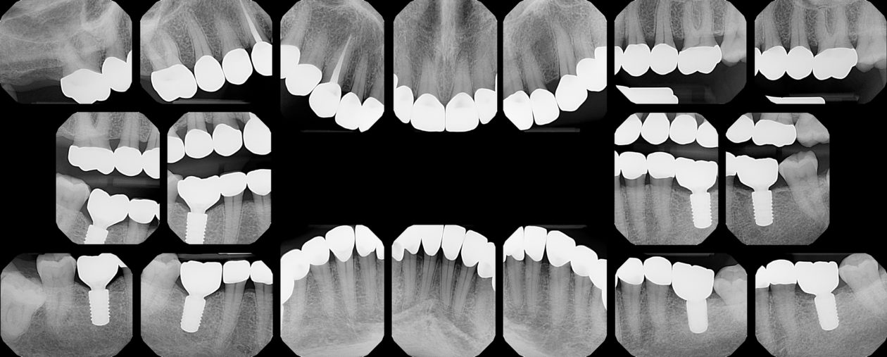
Post-operative FMX

Phase IX: Post-op Evaluation/Radiographs/Photographs

  • Patient returned after one week for further refinement of occlusion.
  • Post-operative radiographs were made.
  • Final post-operative photographs were made.

Phase X: Maintenance

  • Three-month dental prophylaxis schedule.
  • Oral hygiene compliance monitoring.
  • Erosion monitoring.
Definitive tooth display at rest.
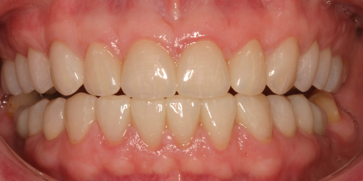
Definitive maximum intercuspation.
Definitive protrusive.
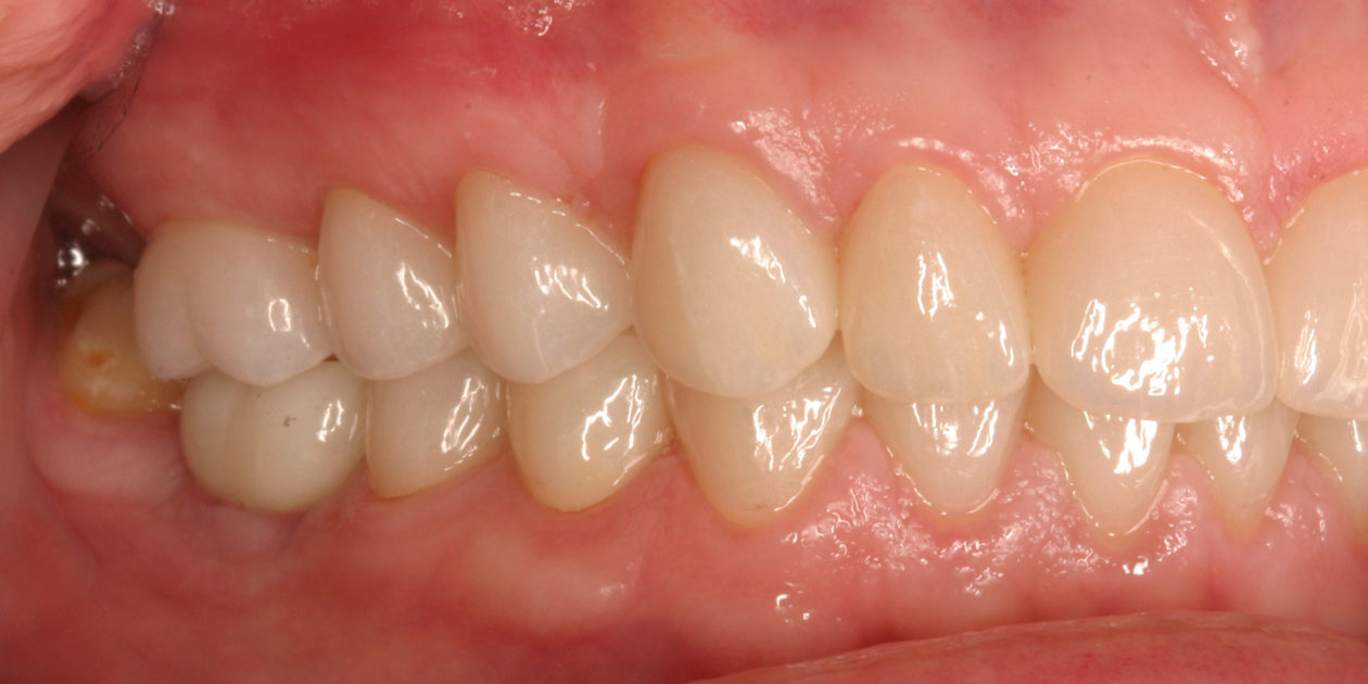
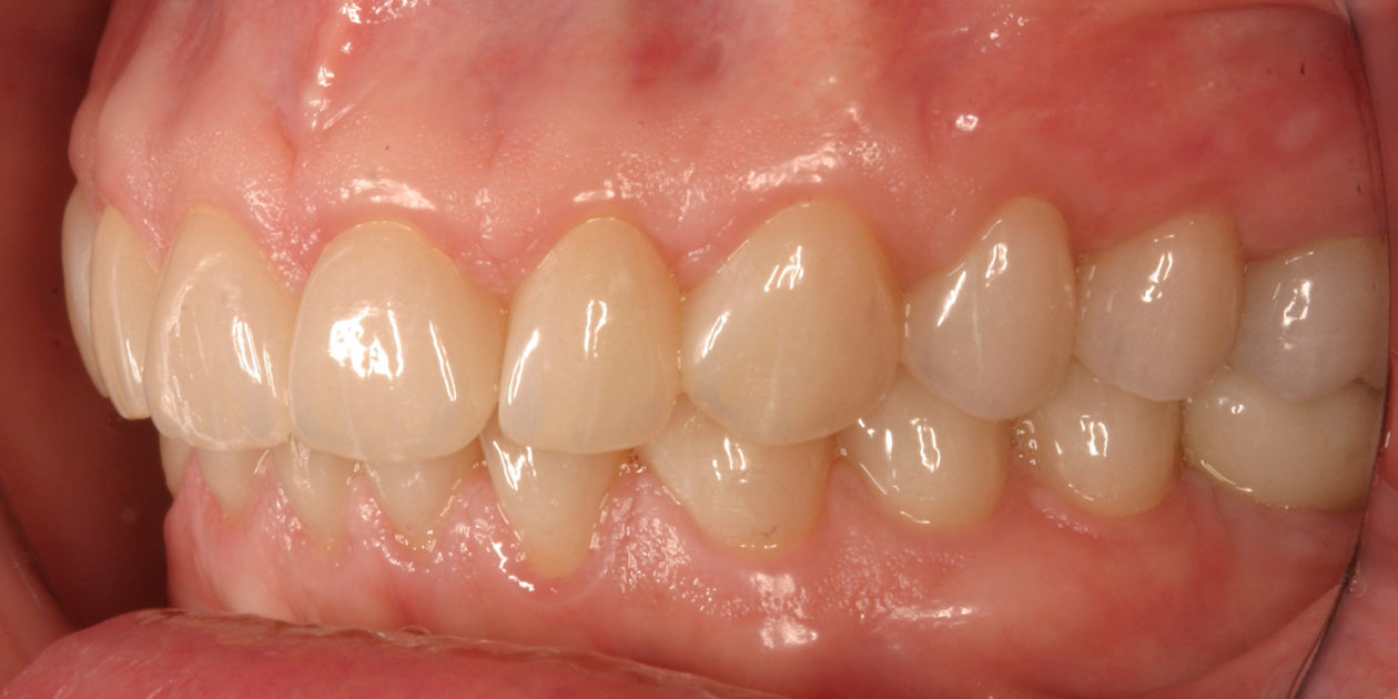
Definitive right lateral view.
Definitive left lateral view.

Commentary

Definitive full face.

The restorative outcome in this case was successful esthetically, functionally and structurally. However, the prognosis regarding the biological aspects of the case remain guarded. The biological aspects of concern in this case include psychology, neuromuscular disorders, and dental erosion.

We achieved psychological success in reducing the patient’s dental phobia. The patient’s eventual comfort and ability to tolerate extensive restorative treatment was achieved. This was possible through three actions: 1) increasing motivation for treatment by creating a vision of the final outcome through simulations, 2) oral medication/nitrous sedation, and increasing dentist trust/confidence through deliberate psychological reassurances, and 3) pain management. As treatment progressed, the patient elected to discontinue oral sedation prior to procedures and required less reassurance at each appointment.

We remain concerned, however, with the long-term biological success of this case due to ongoing bruxism and erosion that contributed to her presenting dental deterioration. The patient’s occlusal/incisal wear pattern at initial presentation reflected an anteriorly directed bruxism pattern. Our restorative plan was modified to include an occlusal scheme designed to distribute functional contacts over as many teeth as possible and allow freedom of movements along the pathways of her former wear patterns. We also modified the material selections to utilize those best able to tolerate increased force levels during parafunction. She has also been fitted with a fully adjusted occlusal orthotic device to be worn while sleeping. However, experience tells us that her potential for parafunctional occlusal trauma remains.

And finally, the causes of the patient’s significant dental erosion at initial presentation remain unclear. We have recommended traditional methods for increasing oral pH (baking soda rinses, pH 9.5 bottled water, and abrasive-free toothpastes). We have also a recommended medical evaluation to rule-out gastro-esophageal reflux disease or any other contributing medical factors.

We will continue to monitor the patient at future appointments with increased attention to these potential threats to her successful restorative outcome.

This case presented an opportunity to evaluate treatment options available to resolve excess dento-alveolar eruption. Orthodontic correction, periodontal therapies, and prosthodontic therapies were considered. We utilized the following analysis to determine the necessary treatment to achieve our esthetic, functional and structural goals.

Clinical Findings

  1. Tooth wear and corresponding length/width disproportion.
  2. Incisal display at repose/rest of negative 2mm.
  3. Gingival display at maximum smile of 3mm.
  4. Vertical axial wall height on palatal of maxillary anterior teeth at 0 to 1mm on a patient with a protrusive bruxism habit.

Goals of Treatment

  1. Improve tooth proportions.
  2. Tooth display at rest + 1mm.
  3. Gingival display 2mm or less.
  4. Gain minimum 2mm ferrule for retentive form.

Treatment Options to Address Dento-Alveolar Extrusion

  1. Comprehensive orthodontic treatment.
  2. Pre-prosthetic crown lengthening.
  3. Both.

Through an effort in structural and functional analysis, a digital diagnostic wax up was developed. This visualization allowed us to determine that the esthetic, structural and functional treatment goals could be met without comprehensive orthodontic treatment.

This analysis confirmed:

  1. Alignment correction of tooth no. 10 could be successfully managed prosthetically.
  2. Gingival margin irregularities of maxillary/mandibular anterior teeth could successfully managed by esthetic crown lengthening on the facial surfaces.
  3. Full coverage prosthetic can be achieved by functional crown lengthening on maxillary teeth. This will also allow for sub-gingival margin placement to protect against the chemical erosion of her dentition.
  4. A desirable occlusal scheme could be developed to mitigate her parafunctional habits.

Dr Gene Ranieri is a periodontist practicing in Valparaiso, Indiana, an adjunct faculty member at Indiana University School of Dentistry, and director of the Dental Study Club of Northwest Indiana branch of the Seattle Study Club.

Dr Michael Drone is a prosthodontist practicing in Valparaiso, Indiana.

Ceramic technicians Aleksandra Polczynski and Jack Polczynski from Visual Dental Arts.