Case 11: Special Report 11

As the evolution of esthetic materials in dentistry continues, clinicians have had to balance strength and beauty: Gold is unbreakable but ugly; feldspathic porcelain is beautiful but weak. Years ago, the introduction of materials such as lithium disilicate somewhat bridged that gap and gave us good esthetics and good strength. With the introduction of zirconia-based materials, we then had great strength and acceptable esthetics. The latest generation of zirconia-based materials has now raised the beauty of zirconia to the level of lithium disilicate. This Special Report showcases the high esthetics that can be achieved with a zirconia-based material in the hands of superior clinicians and lab technicians.

Introduction and Background

A 64-year-old woman—a patient of record since May 1997—presented with a porcelain-fused-to-metal (PFM) bridge spanning teeth nos. 9 to 11 that had been placed by another clinician 15 years prior to becoming the author’s patient. She had never been satisfied with the appearance of the bridge but believed it could not be improved. The patient was seen regularly for hygiene appointments and basic reparative dentistry. Despite expressing interest over the years when discussing esthetic options for replacing the PFM bridge, she never followed through. In August 2017, the patient was retiring and beginning a new chapter in her life. She said she was ready to replace the unesthetic bridge and wanted her other teeth to appear natural and white.

Medical History

The patient was healthy and reported no medical conditions or medications.

Diagnostic Findings

Extraoral/Facial

  • Thin, hypermobile upper lip.
  • Hypermobility of upper lip suppressed in normal smile.

Intraoral/Dental

  • Missing teeth nos. 1, 10, 15, 16, 17.
  • Bleeding on probing exhibited at teeth nos. 3, 14, 19, 30.
  • Asymmetry of length and shape of all anterior teeth.
  • Inconsistent tooth color.
  • Signs of wear and chipping on the incisal edges of the natural anterior dentition.
  • PFM bridge replacing teeth nos. 9–11.

TMJ/Muscles of Mastication

  • Range of motion normal.
  • No deviation on opening.
  • Joint sounds normal.
  • All muscles of mastication exhibit no tenderness to palpation.

Periodontal

  • Oral hygiene good.
  • Periodontal health good.
  • No mobilities.
  • No probing more than 4 mm.
  • Generalized gingival recession of 1 to 3 mm.
Preoperative frontal view of a 64-year-old patient who presented for replacement of old restorations.
Preoperative retracted facial view of the maxillary and mandibular arches. Note multiple colors among the teeth and restorations.

Panoramic Radiograph 2017

Occlusal Notes

CR-CO Discrepancies

  • Equilibration performed 5 years earlier.
  • In the period since equilibration, CR = MIP.
  • Patient has worn a full-coverage acrylic maxillary appliance fabricated in centric relation for 5 years.

Interarch Relationships

  • Class I occlusion with canine rise bilaterally.
  • Less-than-ideal anterior guidance due to irregular incisal edges of mandibular teeth.
  • History of nocturnal bruxism.
  • Visible signs of wear on multiple teeth.

Radiographic Review

  • Root morphology normal, with no pathology.
  • Missing teeth nos. 1, 10, 15, 16, 17.
  • Tooth no. 32 impacted.
  • Lack of adequate bone at site no. 10 to support a dental implant.
Vertical bitewings 2017.

Diagnosis and Prognosis

  • Pre-existing restorations at teeth nos. 9–11 have low esthetic value and less-than-ideal anterior guidance.
  • Prognosis is good given the patient’s good oral hygiene and acceptable occlusion.

Summary of Concerns

  • The chief concern is whether we can meet the patient’s high esthetic demands.
  • The patient declined to have bone grafting surgery, preferring to return to a bridge restoration. She felt she experienced good longevity with her first bridge restoration.
  • We will need to use a high-strength material for the bridge with esthetic matching abilities for use in restorations of other teeth.
  • Can we improve anterior function and guidance?

Proposed Treatment Plan

Driven by the patient’s desire for a more esthetic, natural-looking smile, the treatment plan involved preparing teeth nos. 4–9, 11–13, and 22–27. The maxillary teeth would be restored with a combination of an esthetic, high-strength, medium-translucency multi-zirconia material (IPS e.max ZirCAD MT Multi, Ivoclar Vivadent) and a lithium disilicate material (IPS e.max, Ivoclar Vivadent). The mandibular restorations would be fabricated with a lithium disilicate material only (IPS e.max). Additionally, the case would be completed with a bite splint.

The selected zirconia material would provide the requisite strength for the bridge restoration and enable seamless blending with the proposed single-unit lithium disilicate restorations. The machinable lithium disilicate blocks would impart both monolithic strength and esthetics to the restorations. With a single ceramic block, full-contour restorations can be fabricated, thereby eliminating the challenges associated with layering different materials. However, although layering isn’t required, staining and glazing can be performed to achieve more individualized characterization and blending with other restorations and natural teeth, when necessary.

Active Clinical Treatment

The digital impression scan was overlaid with a full-face photo of the patient to virtually evaluate the case design.
View of the final digital design on screen with the translucency increased, showing the preoperative tooth positions (in blue) through the design.
The definitive restorative design was digitally positioned and shaped to match the provisionals as closely as possible.
View of the full facial smile photo of the patient in the provisional restorations aligned with the scan of the provisionals.

Phase I: Diagnostic Work-up

Full-arch maxillary and mandibular intraoral impression scans and bite registration (Trios, 3Shape) were taken, in addition to a complete series of photographs and radiographs. When integrated with CAD technology in the dental laboratory, intraoral impression scans can be used to create a digital diagnostic wax-up, which serves as a virtual tool for treatment planning and case design. These digital records were forwarded to the laboratory ceramist for use in creating a digital diagnostic design. The ceramist provided a putty matrix based on the digital design for use during fabrication of the chairside provisional restorations.

Phase II: Initial Digital Design and Wax-up

At the laboratory, the impression scans were overlaid with the patient’s full-face photo so that the proposed design could be superimposed with the patient’s face. This essentially enabled a virtual try-in of the design, digital verification that the midline was vertical and the incisal display was level with the face. Additionally, this allowed for an overall evaluation of the esthetic components of the design and provided useful information for planning the tooth preparations.

For example, when viewing the final digital design on-screen with the translucency increased, the preoperative tooth positions (in blue) can be seen through the design. This illustrates the manner in which the buccal corridor will be widened in the premolar area, lengthening the right side and shortening tooth no. 11. This also reveals the need for tissue recontouring at teeth nos. 9 and 10. Further, studying the case in this manner enables the clinician to visualize where more tooth structure will need to be removed and where less reduction will be required in order to achieve final treatment goals.

The diagnostic designs of the maxillary and mandibular restorations were then sent to a 3D printer (FormLab2) for creating printed models which were, in turn, used to create silicone matrices from which the provisional restorations would be fabricated intraorally.

Phase III: Preparation and Provisionalization

Anesthesia was administered and the teeth were prepared for full-coverage crown restorations. Final digital impressions, bite registration scans, and photographs of the preparations were taken. Provisional restorations were made using a dual-curing, nano-technology provisional composite (Luxatemp, DMG).

During the one-week post-preparation appointment, the patient reported that she was doing well and was happy with the provisional restorations. The occlusion was refined, and slight incisal edge changes were made. Intraoral scans and photographs of the provisionals were taken and transferred to the laboratory ceramist for use in initiating the definitive case design.

Special effects were stained onto the zirconia bridge prior to sintering.
View of the zirconia bridge after sintering.

Phase IV: Laboratory Fabrication

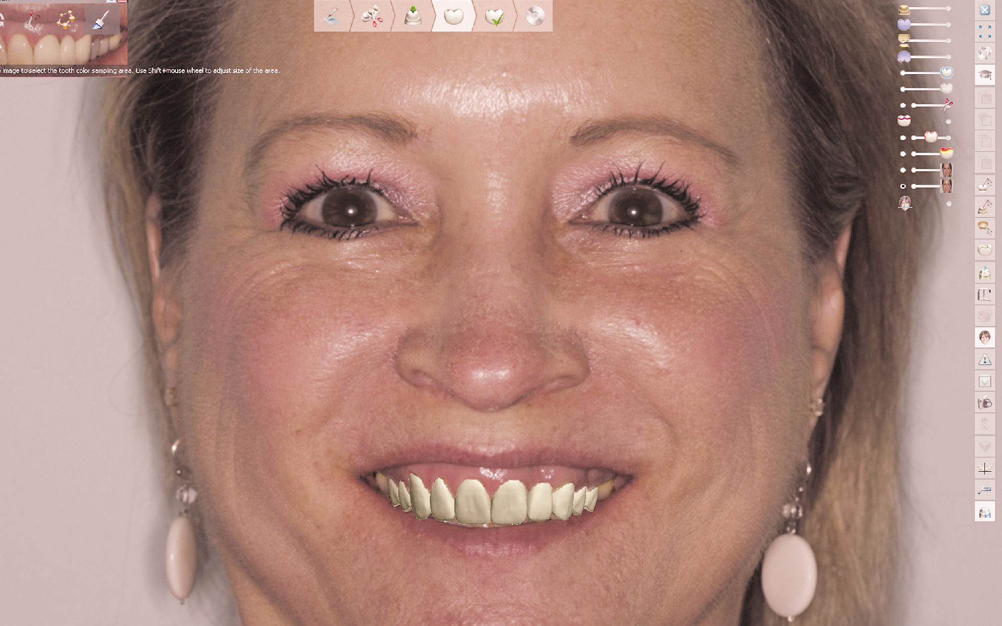
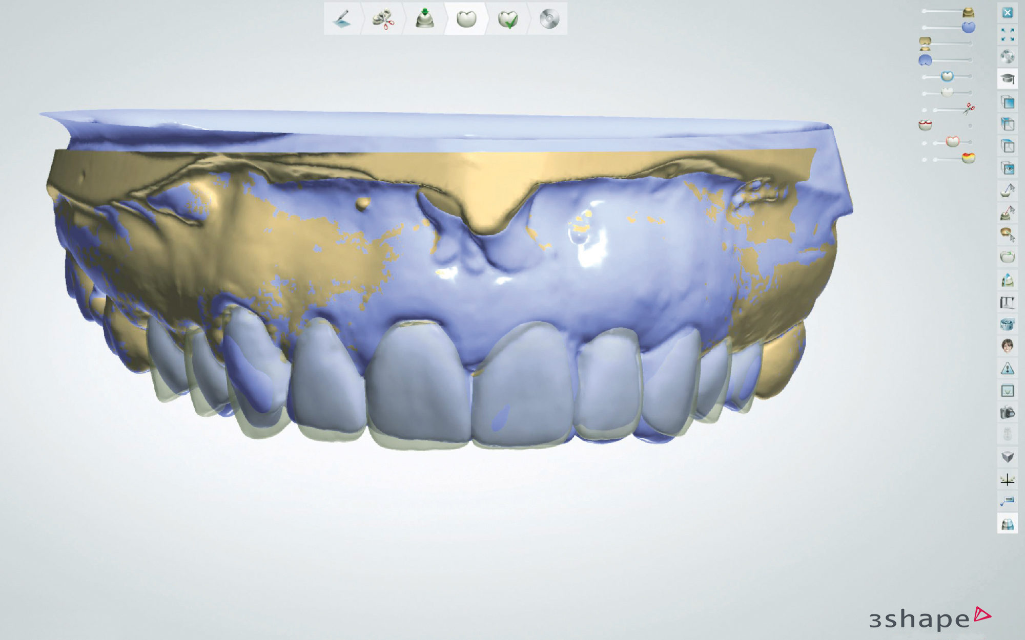
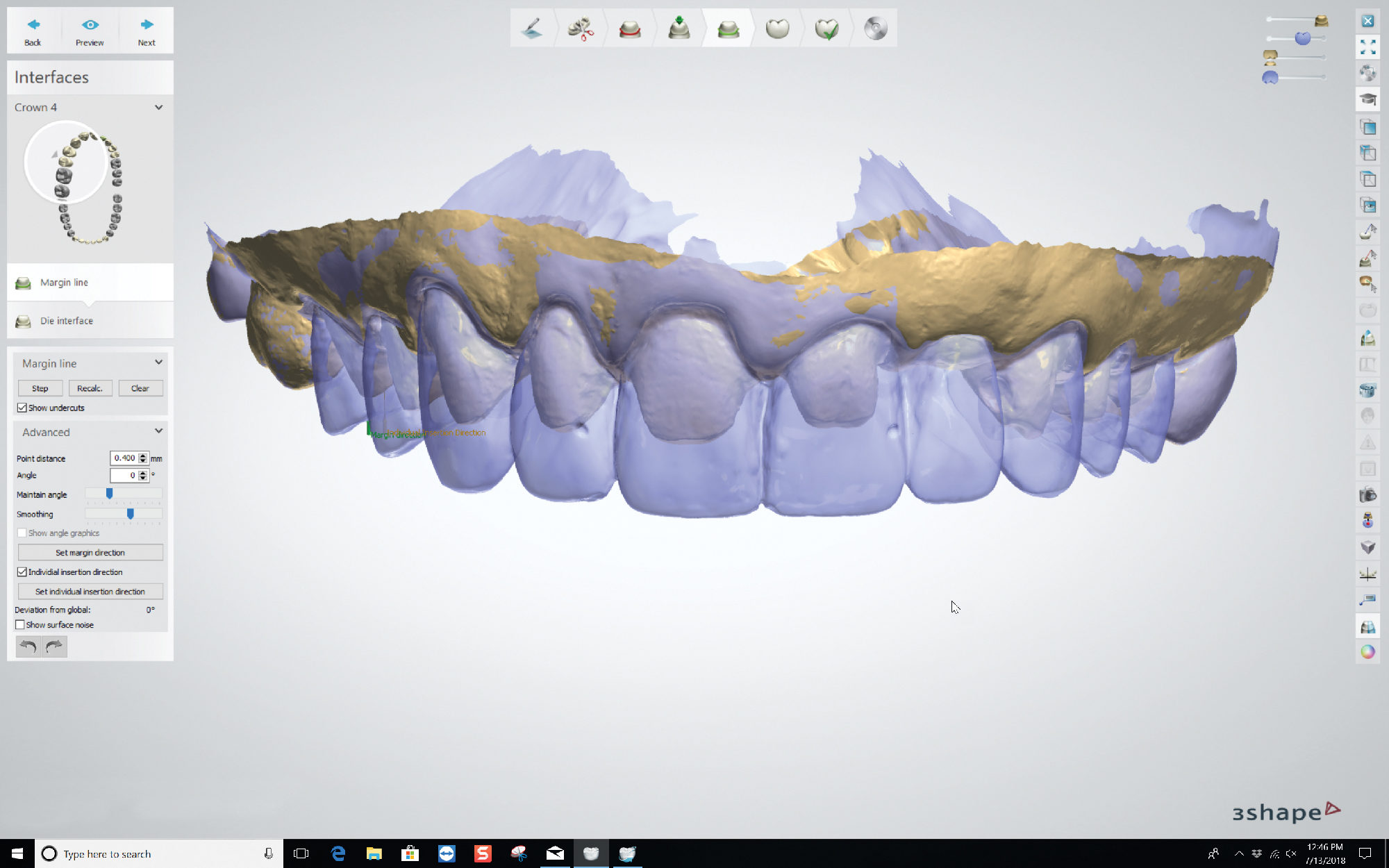
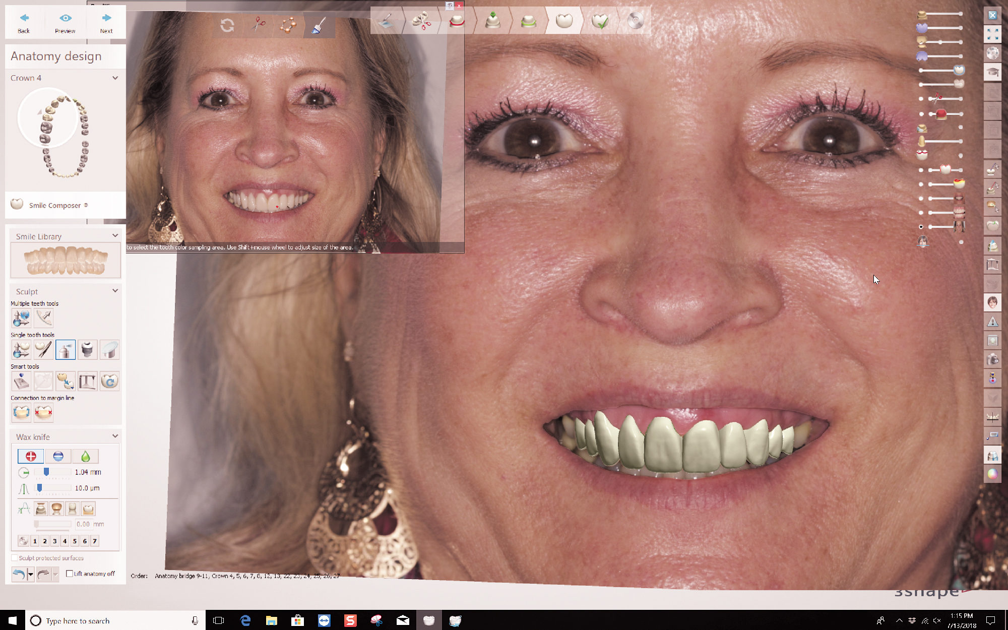
Prior to proceeding with fabrication of the definitive restorations, a full facial analysis of the provisional restorations was completed by aligning the scan of the prepared teeth, the scan of the provisionals, and a full-face smile of the patient in provisionals. The definitive restorations were digitally designed to match the form and function of the provisionals as closely as possible.

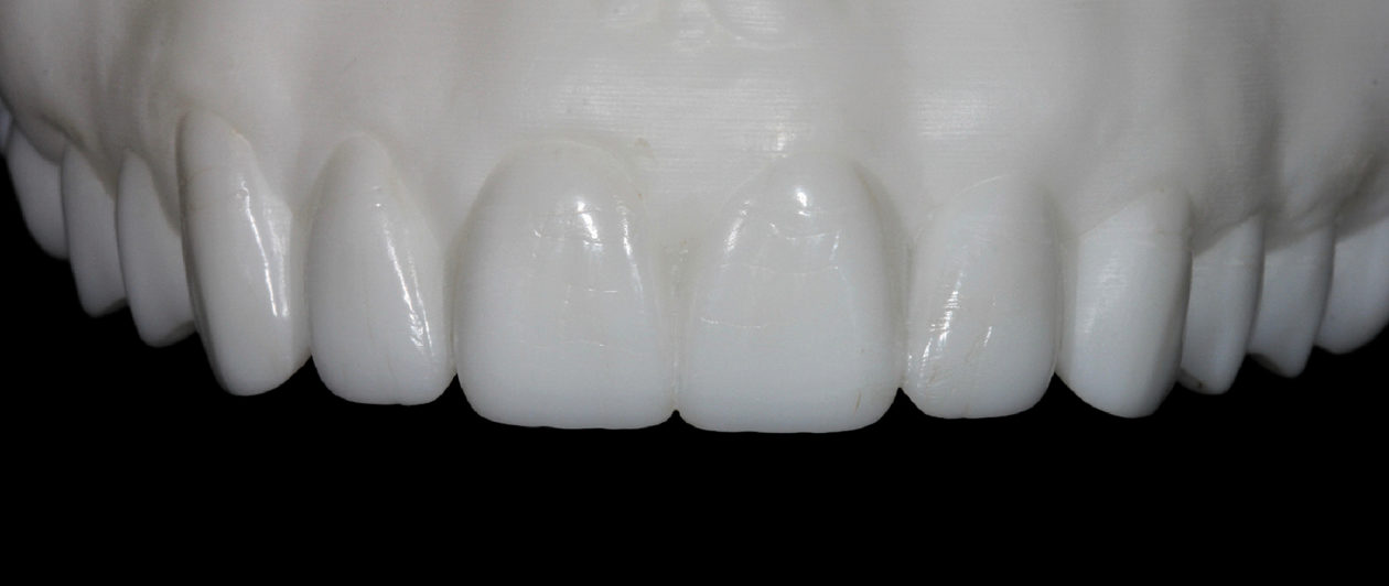
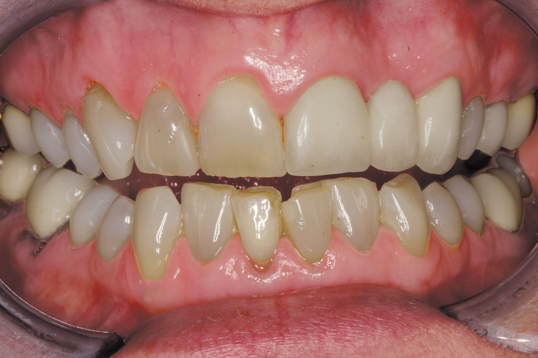
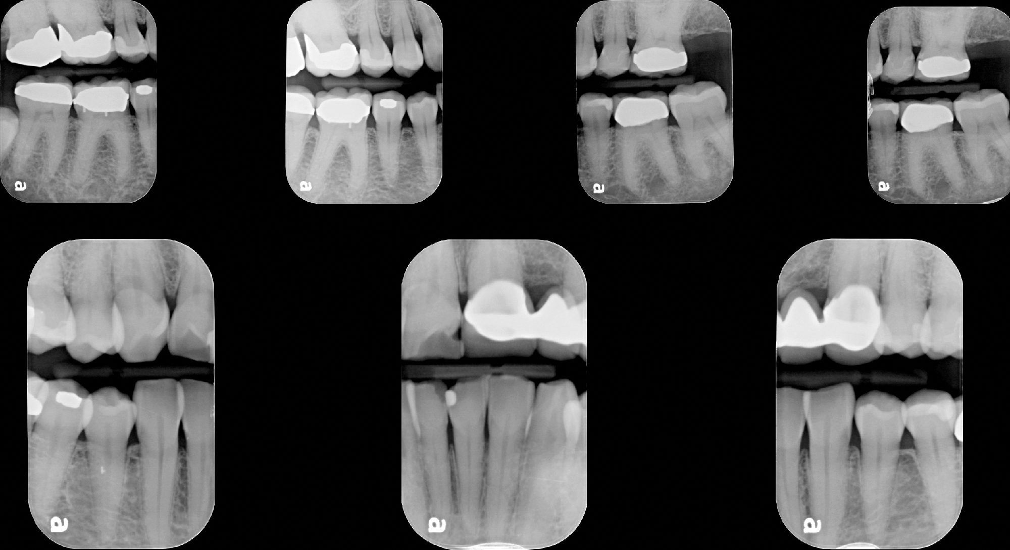
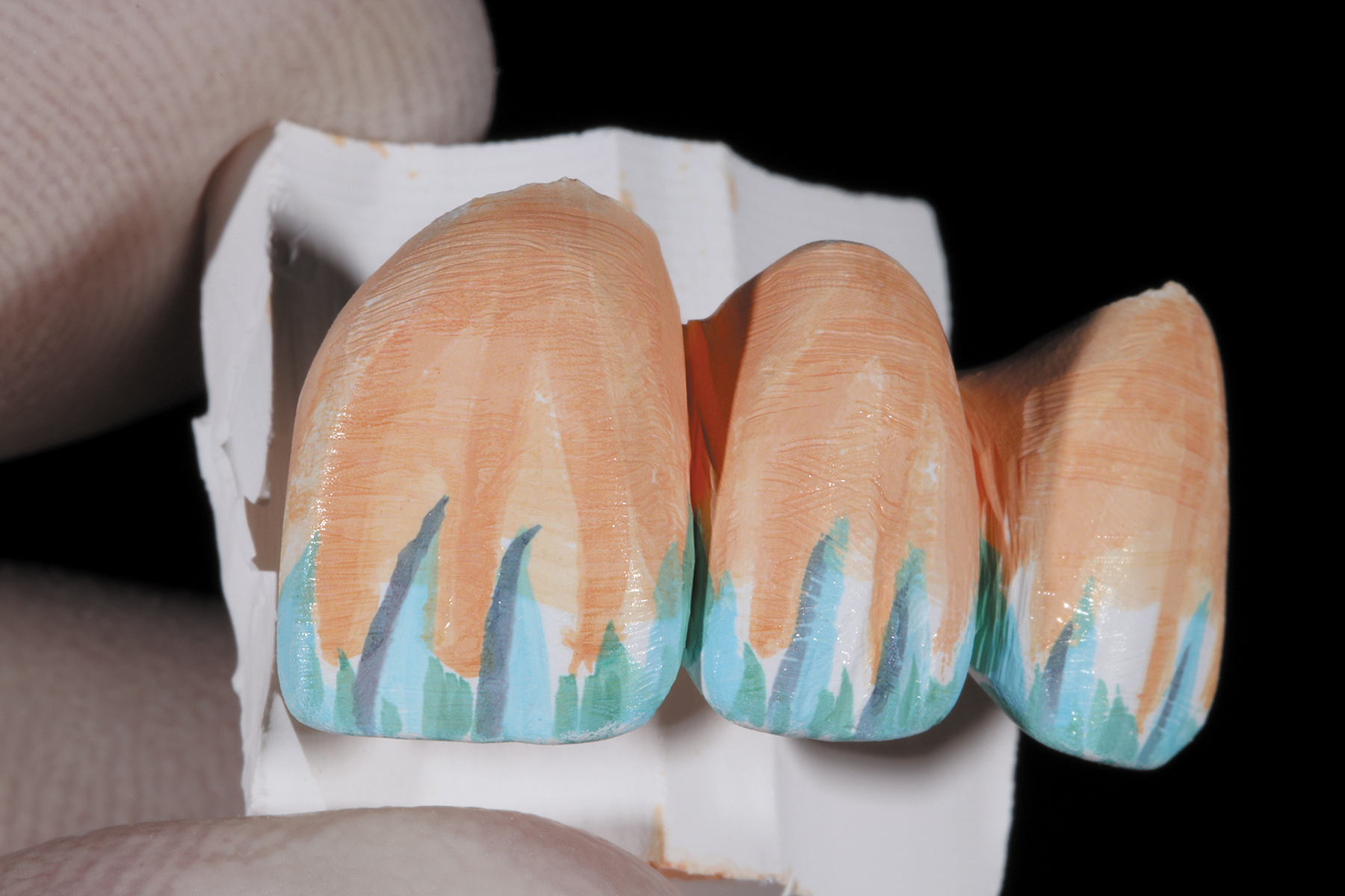
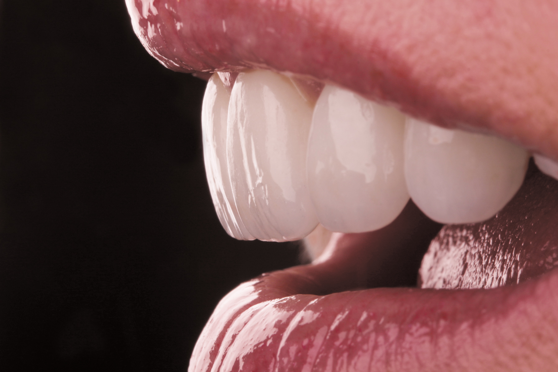
After the MT Multi zirconia bridge was milled, subtle surface morphology was added and special effects were stained into the zirconia prior to sintering. After sintering was complete, the bridge demonstrated a life-like color gradient and surface texture.

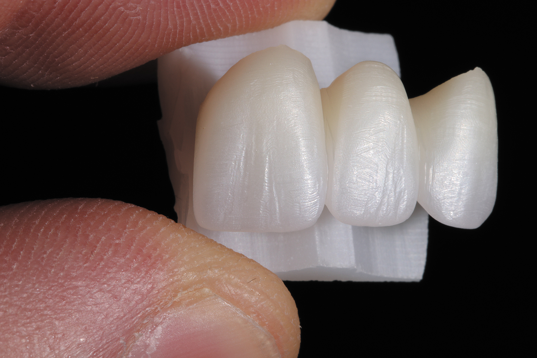
The remaining maxillary anterior zirconia restorations were milled and sintered. Staining and effects (IvoColor Staining, Ivoclar Vivadent) were applied and the color was evaluated to achieve a harmonious blend with the premolars, which were fabricated from a machinable lithium disilicate material (IPS e.max CAD). The zirconia restorations were glazed (IvoColor Glaze, Ivoclar Vivadent), ultimately enhancing the translucency and color gradient of the monolithic zirconia material.

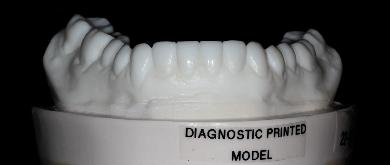
The mandibular anterior restorations were then milled from the machinable lithium disilicate material (IPS e.max CAD) and sintered.

Phase V: Delivery Appointment

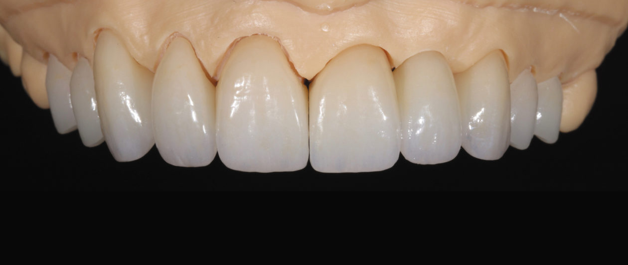
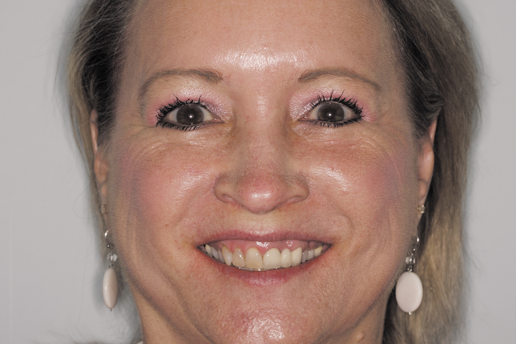
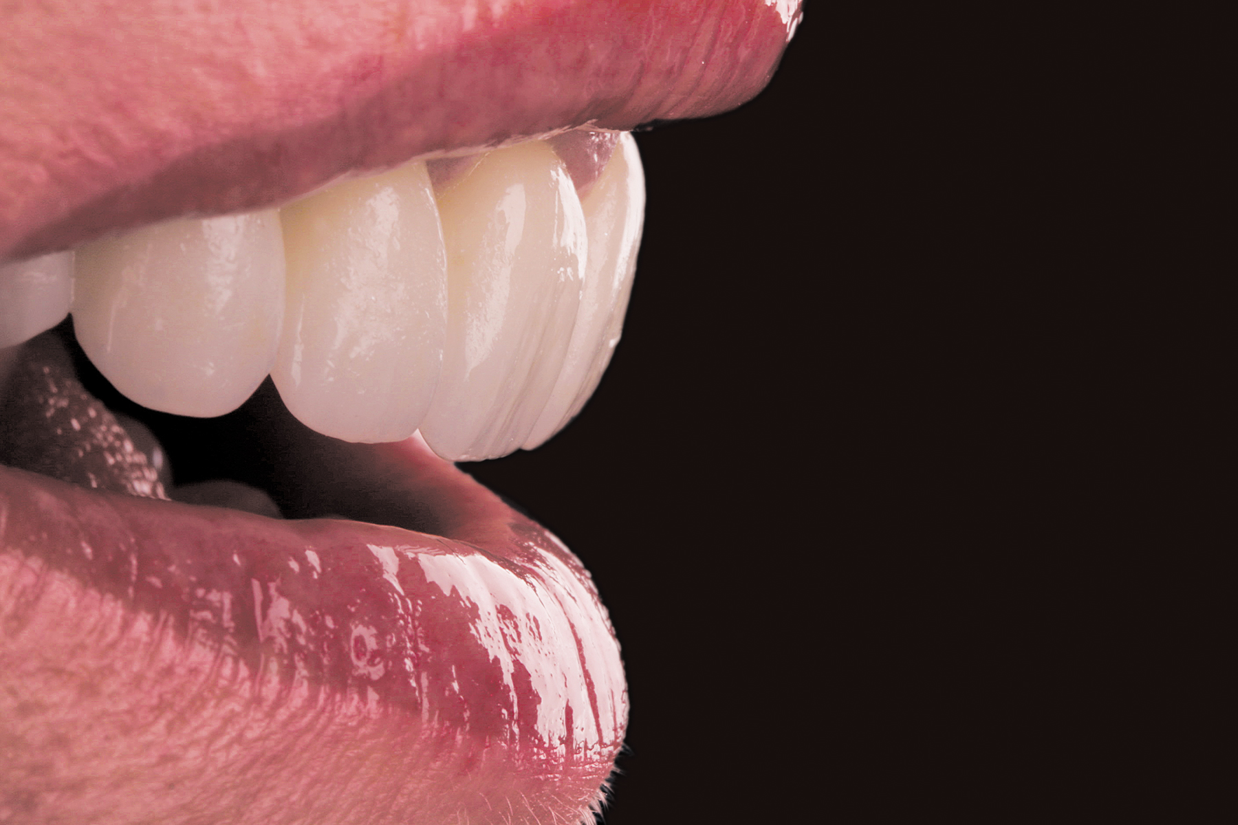
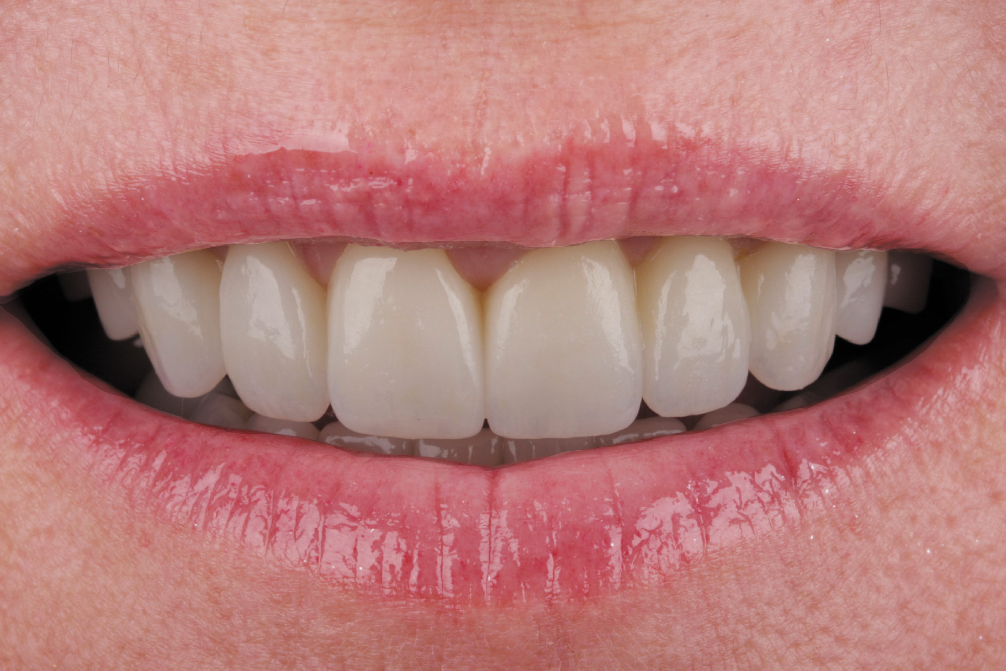
After delivery of anesthetic, the maxillary provisional restorations were removed and the definitive restorations were tried in and adjusted as needed. The patient approved the esthetics by viewing the restorations in multiple lighting situations and on photographs.

Prior to cementation, the intaglio surfaces of the zirconia restorations were cleaned with a universal cleaning paste (Ivoclean, Ivoclar Vivadent) and then rinsed and dried. Next, a self-adhesive, self-curing resin cement (SpeedCEM Plus, Ivoclar Vivadent) was loaded into the restorations. They were then seated on the preparations and the margins were tack-cured. Excess cement was easily and predictably removed.

The maxillary premolar and mandibular anterior lithium disilicate restorations were cleaned with the universal cleaning paste, rinsed, and dried. A universal primer for conditioning restoration surfaces (Monobond Plus, Ivoclar Vivadent) was applied to the intaglio surfaces of the restorations for 60 seconds, and the excess was removed. All enamel and dentin surfaces were etched with phosphoric acid for 15 seconds and thoroughly rinsed. A single-component light-cured adhesive (Adhese, Ivoclar Vivadent) was applied to the preparations, thinned, and light-cured. A neutral shade of a light- and dual-curing luting composite (Variolink Esthetic, Ivoclar Vivadent) was loaded into the restorations, and they were seated on the preparations. The restorations were tack-cured at the margins, excess cement was removed, the interproximal areas were flossed, and the restorations underwent a final cure. The occlusion was then checked and refined.

The patient returned after 3 days for further refinement of the occlusion. Records were taken for fabrication of a new bite splint, and final postoperative photographs were taken.

Right lateral postoperative view of the definitive restorations.
Left lateral postoperative view of the definitive restorations.
Close-up of the postoperative facial view.
Postoperative frontal view.

Commentary

Digital workflows that maximize the use of high-strength millable materials—such as those used for the restorations in this case—contribute to greater efficiency, accuracy, and even creative artistry. The selected materials offer versatility in terms of functional strength while simultaneously enabling harmonious and seamless esthetics across all of the different restorations. The MT Multi zirconia material, in particular, proved to be a very esthetic option for the three-unit bridge in this anterior case.

For this patient, digital processes also equated to a more convenient and precise treatment. The patient was very pleased with the esthetics and function of her new restorations. Our only critique of the case was that the patient was not interested in—and therefore declined—alteration of the gingival heights to improve symmetry.

Franklin Shull, DMD, is a faculty member at Palmetto Health Richland Department of Dentistry and the Medical College of Georgia School of Dentistry. He also serves on the teaching faculty at the LD Pankey Institute in Key Biscayne, Florida. A published author in many dental journals who lectures throughout the US on esthetics, dental materials, and photography, he recently contributed to a textbook on comprehensive dentistry. A fellow of the Academy of General Dentistry, Dr Shull is also a past president of both the Greater Columbia Dental Association and the SC Academy of General Dentistry.

Matt Roberts, CDT, founded CMR Dental Laboratory in 1979, where he elevates the quality of restorative treatments received by patients by creating an interactive working relationship among dentists, ceramists, and specialists. An accredited member of the American Academy of Cosmetic Dentistry, he lectures nationally and internationally and has worked with many of the country’s leading clinicians. Mr Roberts is also the founder of Team Aesthetic Seminars and conducts advanced-level training classes for dentists and ceramists.